News
Autophagie-Anwendungen
Yoshinori Ohsumi, honorary professor at the Tokyo Institute of Technology, was awarded the 2016 Nobel Prize in Physiology or Medicine for his discovery of autophagy (autophagocytosis). His research into autophagy started with observing yeasts using an optical microscope. Interest in autophagy has expanded from yeast cells to mammalian and other type of animal cells through the work of many researchers, and autophagy is currently the focus of many research groups worldwide.
Autophagy research ranges from fundamental research, such as understanding how autophagy works on a molecular level and what roles it plays in living organisms, to clinical applications, such as examining how autophagy is associated with Alzheimer’s disease and other neurological diseases. Olympus’ biological microscopes have contributed to this leading-edge autophagy research.
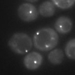
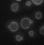
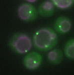
Fluorescence was used to observe the autophagy-related protein Atg17 and vacuole membranes in budding yeasts (Atg17 mutant). Researchers used oblique illumination with a 150X high-magnification TIRF objective lens and the IX3 inverted microscope.
|
|
|
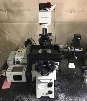
A TIRF microscope in Dr. Ohsumi’s lab at the Tokyo Institute of Technology
Image data courtesy of Hayashi Yamamoto and Yoshinori Ohsumi, Frontier Research Center, Tokyo Institute of Technology
Reference:
Yamamoto H, Fujioka Y, Suzuki SW, Noshiro D, Suzuki H, Kondo-Kakuta C, Kimura Y, Hirano H, Ando T, Noda NN, Ohsumi Y. The Intrinsically Disordered Protein Atg13 Mediates Supramolecular Assembly of Autophagy Initiation Complexes. Dev Cell. 2016 Jul 11;38(1):86-99.
Related product: UAPON150XOTIRF Objective
Simultaneous three-color fluorescence observation of organelles and autophagosomes in COS7 cells using a laser scanning microscope based on a fully-motorized IX series inverted microscope. This setup enables three-color simultaneous fluorescence observation.
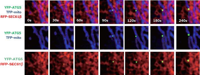
The world’s first dynamic observation of autophagosomes forming from net-like mitochondria (RFP-SEC61β, red) and endoplasmic reticulum (TFP-mito, blue) contact sites.
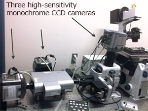 A conventional simultaneous three-color fluorescence observation system | 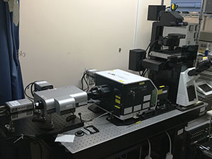 Our current simultaneous three-color fluorescence observation system |
| Live-cell imaging microscope system in Dr. Yoshimori’s lab | |
The picture on the left shows the simultaneous three-color fluorescence imaging system used to capture images that were published in Nature magazine. This system is based on a fully-motorized version of an IX81 inverted microscope. The picture on the right is an upgraded version based on the fully-motorized IX83 microscope.
Image data courtesy of
Maho Hamasaki and Tamotsu Yoshimori, Department of Genetics, Osaka University Graduate School of Medicine
Reference:
Maho Hamasaki, Nobumichi Furuta, Atsushi Matsuda, Akiko Nezu, Akitsugu Yamamoto, Naonobu Fujita, Hiroko Oomori, Takeshi Noda, Tokuko Haraguchi, Yasushi Hiraoka, Atsuo Amano, & Tamotsu Yoshimori.
Autophagosomes form at ER-mitochondria contact sites.
Nature. 2013 Mar 21;495(7441):389-93. doi: 10.1038/nature11910. Epub 2013 Mar 3.
Related products: Fully-Motorized, Automated IX83 Inverted Microscope
Live imaging of the autophagy-related protein LS3 in the fertilized eggs of a GFP-LC3-RFP-LC3 ΔG transgenic zebrafish. Researchers used a 30X silicone immersion objective (UPLSAPO30XS) and a FLUOVIEW laser scanning confocal microscope.
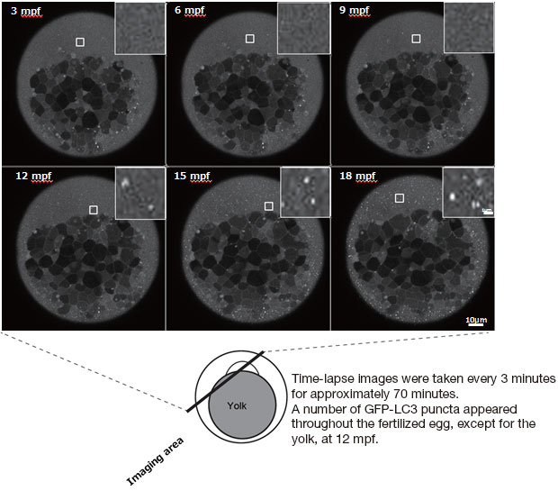
Image data courtesy of
Hideaki Morishita and Noboru Mizushima, Department of Biochemistry and Molecular Biology,
Graduate School and Faculty of Medicine, The University of Tokyo
Reference:
Takeshi Kaizuka, Hideaki Morishita, Yutaro Hama, Satoshi Tsukamoto, Takahide Matsui, Yuichiro Toyota, Akihiko Kodama, Tomoaki Ishihara, Tohru Mizushima, Noboru Mizushima
An Autophagic Flux Probe that Releases an Internal Control.
Mol Cell. 2016 Oct 25. pii: S1097-2765(16)30589-5. doi: 10.1016/j.molcel.2016.09.037. [Epub ahead of print]
Related products: FV3000 Laser Scanning Confocal Microscope and UPLSAPO30XS Silicone Immersion Objective
Loaclization analysis of a soluble N-ethylmaleimide-sensitive factor attachment protein receptor (SNARE) that causes membrane fusion in autophagy-related ATG3 knockout MEF cells under starvation conditions (one hour after starvation).
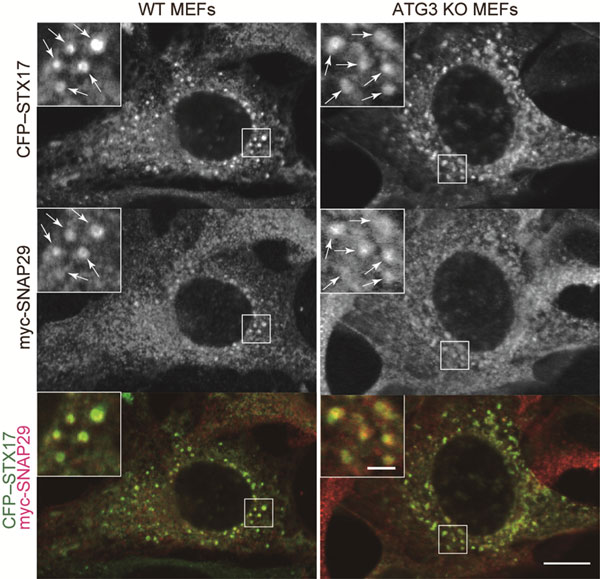
Colocalization of SNARE protein SECFP-STX17 and myc-SNAP29 was observed (white arrow).
Wild type MEF (left) and ATG3 knockout MEF (right)
Scale bar = 10 μm; scale bar in the close-up image within the white frame = 2 μm
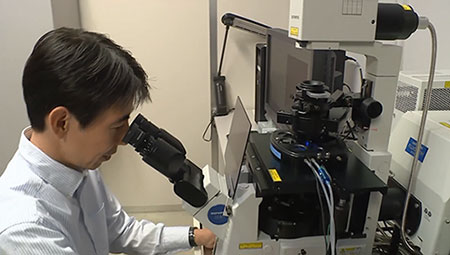
An FV1000 confocal microscope in Dr. Mizushima's lab at theUniversity of Tokyo
Image data courtesy of Kotaro Tsuboyama, Ikuko Honda and Noboru Mizushima, Department of Biochemistry and Molecular Biology, Graduate School and Faculty of Medicine, The University of Tokyo
Reference:
Kotaro Tsuboyama, Ikuko Koyama-Honda, Yuriko Sakamaki, Masato Koike, Hideaki Morishita, Noboru Mizushima.
The ATG conjugation systems are important for degradation of the inner autophagosomal membrane.
Science 20 Oct 2016: DOI: 10.1126/science.aaf6136.
Related products: FV3000 Laser Scanning Confocal Microscope
Researchers conducted super resolution live imaging of autophagosome membranes in mouse fibroblasts using the SD-OSR super resolution microscope system and a 100X silicone immersion objective lens (UPLSAPO100XS).
SD-OSR (Left side of movie)
| Confocal (Right side of movie)
|
Super resolution images of GFP-p62 in mouse fibroblasts that were captured for 35 minutes continuously (left). Autophagosomes can be observed as rings (right) that are clearer than those produced using conventional confocal imaging.
Image data courtesy of Satoshi Waguri,
Department of Anatomy and Histology, Fukushima Medical University
Related product: SD-OSR Spinning Disk Super Resolution System
Because the SD-OSR is special order basis product, please contact us for the detail.
The FV-OSR software enables super resolution imaging using the FV3000 microscope. Researchers used this system to capture images of the WIPI2 protein that initiates autophagosome formation (AlexaFluor488) and the outer mitochondrial membrane protein TOM20 (AlexaFluor568) in MEF fixed with PFA after autophagy induction (1.5 hours of growth in a culture media without amino acid).
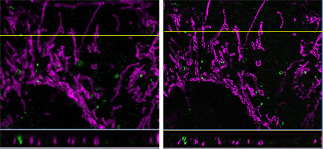
These images show a horizontal section of a Z-slice stack image. The picture on the right is enhanced using cellSens® Dimension software. Super resolution XYZ images were processed with the software’s 3D deconvolution function, resulting in enhanced Z resolution.
Image data courtesy of Ikuko Honda and Noboru Mizushima, Department of Biochemistry and Molecular Biology, Graduate School and Faculty of Medicine, the University of Tokyo
Related product: FV3000 Laser Scanning Confocal Microscope and FV-OSR Super Resolution Software
Contributions to Autophagy Research
Our biological microscopes have contributed to leading-edge research into autophagy spanning many fields ranging from basic medicine to studies of viruses, immunity, and plants. The following references all used Olympus microscopes as part of their research.
Takeshi Kaizuka, Hideaki Morishita, Yutaro Hama, Satoshi Tsukamoto, Takahide Matsui, Yuichiro Toyota, Akihiko Kodama, Tomoaki Ishihara, Tohru Mizushima, Noboru Mizushima
An Autophagic Flux Probe that Releases an Internal Control.
Mol Cell. 2016 Oct 25. pii: S1097-2765(16)30589-5. doi: 10.1016/j.molcel.2016.09.037. [Epub ahead of print]
Related products:UPLSAPO30XS Silicone Immersion Objective
Kotaro Tsuboyama, Ikuko Koyama-Honda, Yuriko Sakamaki, Masato Koike, Hideaki Morishita, Noboru Mizushima.
The ATG conjugation systems are important for degradation of the inner autophagosomal membrane.
Science 20 Oct 2016: DOI: 10.1126/science.aaf6136.
Related products:FV3000 Laser Scanning Confocal Microscope
Kenta Imai, Feike Hao, Naonobu Fujita, Yasuhiro Tsuji, Yukako Oe, Yasuhiro Araki, Maho Hamasaki, Takeshi Noda, Tamotsu Yoshimori.
J Cell Sci. 2016 Oct 15;129(20):3781-3791. Epub 2016 Sep 1.
Related products:FV3000 Laser Scanning Confocal Microscope
Kosaku Shinoda, Yutaka Hasegawa, Kenji Ikeda, Haemin Hong, Qianqian Kang, Yangyu Yang, Rushika M. Perera, Jayanta Debnath, Shingo Kajimura.
Beige Adipocyte Maintenance Is Regulated by Autophagy-Induced Mitochondrial Clearance Svetlana Altshuler-Keylin.
Cell Metab. 2016 Sep 13;24(3):402-19. doi: 10.1016/j.cmet.2016.08.002. Epub 2016 Aug 25.
Related products:MVX10 Macro Zoom Microscope
Junya Hasegawa, Ryo Iwamoto, Takanobu Otomo, Akiko Nezu, Maho Hamasaki, Tamotsu Yoshimori.
Autophagosome–lysosome fusion in neurons requires INPP5E, a protein associated with Joubert syndrome.
EMBO J. 2016 Sep 1;35(17):1853-67. doi: 10.15252/embj.201593148. Epub 2016 Jun 23.
Related products:FV3000 Laser Scanning Confocal Microscope
Christopher P Webster, Emma F Smith, Claudia S Bauer, Annekathrin Moller, Guillaume M Hautbergue, Laura Ferraiuolo, Monika A Myszczynska, Adrian Higginbottom, Matthew J Walsh, Alexander J Whitworth, Brian K Kaspar, Kathrin Meyer, Pamela J Shaw, Andrew J Grierson, Kurt J De Vos.
The C9orf72 protein interacts with Rab1a and the ULK1 complex to regulate initiation of autophagy.
EMBO J. 2016 Aug 1;35(15):1656-76. doi: 10.15252/embj.201694401. Epub 2016 Jun 22.
Related products:IX83 Fully Motorized, Automated Inverted Microscope
Hayashi Yamamoto, Yuko Fujioka, Sho W. Suzuki, Daisuke Noshiro, Hironori Suzuki, Chika Kondo-Kakuta, Yayoi Kimura, Hisashi Hirano, Toshio Ando, Nobuo N. Noda & Yoshinori Ohsumi.
The Intrinsically Disordered Protein Atg13 Mediates Supramolecular Assembly of Autophagy Initiation Complexes.
Dev Cell. 2016 Jul 11;38(1):86-99.
Related products:UAPON150XOTIRF Objective
Chantal Sellier, Maria‐Letizia Campanari, Camille Julie Corbier, Angeline Gaucherot, Isabelle Kolb‐Cheynel, Mustapha Oulad‐Abdelghani, Frank Ruffenach, Adeline Page, Sorana Ciura, Edor Kabashi, Nicolas Charlet‐Berguerand.
Loss of C9ORF72 impairs autophagy and synergizes with polyQ Ataxin‐2 to induce motor neuron dysfunction and cell death.
EMBO J. 2016 Jun 15;35(12):1276-97. doi: 10.15252/embj.201593350. Epub 2016 Apr 21.
Related products:IX83 Fully Motorized, Automated Inverted Microscope
Jennifer Martinez, Larissa D. Cunha, Sunmin Park, Mao Yang, Qun Lu, Robert Orchard, Quan-Zhen Li, Mei Yan, Laura Janke, Cliff Guy, Andreas Linkermann, Herbert W. Virgin & Douglas R. Green.
Noncanonical autophagy inhibits the autoinflammatory, lupus-like response to dying cells.
Nature. 2016 May 5;533(7601):115-9. doi: 10.1038/nature17950. Epub 2016 Apr 20.
Related products:BX53 Semi-Motorized Fluorescence Microscope
Shuo Wang, Pengyan Xia, Guanling Huang, Pingping Zhu, Jing Liu, Buqing Ye, Ying Du & Zusen Fan.
FoxO1-mediated autophagy is required for NK cell development and innate immunity.
Nat Commun. 2016 Mar 24;7:11023. doi: 10.1038/ncomms11023.
Related products:FV3000 Laser Scanning Confocal Microscope
Jiwon Jang, Yidi Wang, Matthew A. Lalli, Elmer Guzman, Sirie E. Godshalk, Hongjun Zhou, Kenneth S. Kosik.
Primary Cilium-Autophagy-Nrf2 (PAN) Axis Activation Commits Human Embryonic Stem Cells to a Neuroectoderm Fate.
Cell. 2016 Apr 7;165(2):410-20. doi: 10.1016/j.cell.2016.02.014. Epub 2016 Mar 24.
Related products:IX73 Inverted Microscope System for Advanced Live Cell Imaging
Takeshi Yamamoto, Yoshitsugu Takabatake, Tomonori Kimura, Atsushi Takahashi, Tomoko Namba, Jun Matsuda, Satoshi Minami, Jun-ya Kaimori, Isao Matsui, Harumi Kitamura, Taiji Matsusaka, Fumio Niimura, Motoko Yanagita, Yoshitaka Isaka & Hiromi Rakugi.
Time-dependent dysregulation of autophagy: implications in aging and mitochondrial homeostasis in the kidney proximal tubule.
Autophagy. 2016 May 3;12(5):801-13. doi: 10.1080/15548627.2016.1159376. Epub 2016 Mar 17.
Related products:FV3000 Laser Scanning Confocal Microscope
Chenran Wang, Song Chen, Syn Yeo, Gizem Karsli-Uzunbas, Eileen White, Noboru Mizushima, Herbert W. Virgin, and Jun-Lin Guan.
Elevated p62/SQSTM1 determines the fate of autophagy-deficient neural stem cells by increasing superoxide.
J Cell Biol. 2016 Feb 29;212(5):545-60. doi: 10.1083/jcb.201507023.
Related products:DP74 Digital Microscope Camera
Satoshi Hirano, Takefumi Uemura, Hiromichi Annoh, Naonobu Fujita, Satoshi Waguri, Takashi Itoh & Mitsunori Fukuda.
Differing susceptibility to autophagic degradation of two LC3-binding proteins: SQSTM1/p62 and TBC1D25/OATL1.
Autophagy. 2016;12(2):312-26. doi: 10.1080/15548627.2015.1124223.
Related products:UAPON100XOTIRF Objective
Qun Lu, Christine C. Yokoyama, Jesse W. Williams, Megan T. Baldridge, Xiaohua Jin, Brittany DesRochers, Traci Bricker, Craig B. Wilen, Juhi Bagaitkar, Ekaterina Loginicheva, Alexey Sergushichev, Darren Kreamalmeyer, Brian C. Keller, Yan Zhao, Amal Kambal, Douglas R. Green, Jennifer Martinez, Mary C. Dinauer, Michael J. Holtzman, Erika C. Crouch, Wandy Beatty, Adrianus C.M. Boon, Hong Zhang, Gwendalyn J. Randolph, Maxim N. Artyomov, Herbert W. Virgin.
Homeostatic Control of Innate Lung Inflammation by Vici Syndrome Gene Epg5 and Additional Autophagy Genes Promotes Influenza Pathogenesis.
Cell Host Microbe. 2016 Jan 13;19(1):102-13. doi: 10.1016/j.chom.2015.12.011.
Related products:BX53 Semi-Motorized Fluorescence Microscope
Yurong Li, Mehdi Kabbage, Wende Liu, and Martin B Dickman.
Aspartyl protease mediated cleavage of AtBAG6 is necessary for autophagy and fungal resistance in plants.
Plant Cell. 2016 Jan;28(1):233-47. doi: 10.1105/tpc.15.00626. Epub 2016 Jan 6.
Related products:FV3000 Laser Scanning Confocal Microscope
Not Available in Your Country
Sorry, this page is not
available in your country.
