Ask the Experts
Willkommen auf der Seite von Ask the Experts, dem Portal für Fragen zu Mikroskopie im Bereich der Biowissenschaften. Nehmen Sie an Live-Webinaren teil, um aktuelle Themen zu diskutieren, oder kontaktieren Sie unsere Experten bei Fragen und Herausforderungen jederzeit. Haben Sie ein Webinar verpasst? Kein Problem, Sie können es jederzeit wieder anhören. Haben Sie ein Thema, das Sie diskutieren möchten? Nutzen Sie unser Feedback-Formular oder wenden Sie sich direkt an unsere Experten.
Live-Webinare | Online-Webinare | Experten

Investigating Spheroid Architecture Using the FV3000 Confocal Microscope
Experten
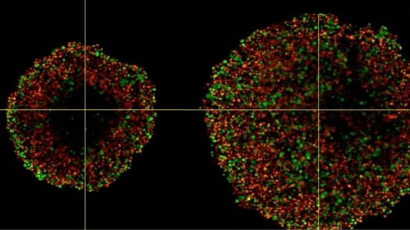
Phenotypic tumour heterogeneity arising due to differentially cycling cell populations has been implicated in increased therapy resistance. This phenomenon cannot be assessed in adherent cell culture, where microenvironmental conditions are homogeneous. Thus, we utilise melanoma spheroids to model the 3D tumour microenvironment including the extracellular matrix (ECM) and study spheroid structure, necrotic region, individual cell arrangement within and gene expression patterns. We achieve this by exploiting the fluorescence ubiquitination cell cycle indicator (FUCCI) system to monitor cell cycle stages as a surrogate marker for phenotypic tumour heterogeneity, tissue clearing and confocal microscopy using FV3000.

Prostate Cell Lineage Hierarchy and Plasticity
Experten
Prostate cancer is one of the most common cancers worldwide and also the second leading cause of cancer-related death in males in Western countries. Although the majority of human primary prostate cancers have a luminal phenotype, both basal cells and luminal cells can serve as cellular origins of prostate cancer in model systems. However, the stem cell-like plasticity of defined prostate epithelial cells and the cellular origin of prostate cancer under physiological conditions have not been identified. Recently, prostate basal and luminal cell populations were both shown to be self-sustaining, and both cell types could initiate prostate cancer. However, the oncogenic transformation of basal cells requires basal to luminal cell transition. In addition, luminal cells were shown to have greater tendency to be the cells of origin for prostate cancer in some contexts.

3D Segmentation for Fluorescence Images: From Qualitative to Quantitative
Experten
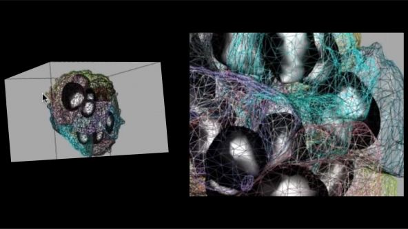
Cells are 3D functional elements in biology science and they are actively moving to perform their functions. Collective cell migration is appreciated as an important model for the understanding of the mechanism governing the cell movement in Vivo and in Vitro. It is a highly kinetic process involved in immune response, wound healing, tissue development and cancer metastasis. Recent decades have seen the fast development of various optical imaging techniques with excellent spatial-temporal resolution, dimensionality and scale. The generation of novel probes have also allowed us to acquire the movies of migrating cells with specific proteins/molecules. However, we lack of advanced solution to analyse such high-content and highly dynamic images/videos.

An In Vitro System for Evaluating Anticancer Drugs Using Patient-Derived Tumor Organoids
Experten
Patient-derived tumor organoids (PDOs) represent a promising preclinical cancer model that better replicates disease, compared with traditional cell culture models. We have established a novel series of patient-derived tumor organoids (PDOs) from various types of tumor tissues from the Fukushima Translational Research Project, which are designated as Fukushima (F)-PDOs. F-PDOs could be cultured for >6 months and formed cell clusters with similar morphologies to their source tumors. Comparative histological and comprehensive gene-expression analyses also demonstrated that the characteristics of PDOs were similar to those of their source tumors, even following long-term expansion in culture. In addition, suitable high-throughput assay systems were constructed for each F-PDO in 96- and 384-well plate formats.

Study the Function of Stromal Cells through Intestinal Organoid Co-Culture Technology
Experten
For a long period of time, intestinal mesenchymal stromal cells have been considered as a relatively simple and homogeneous group of cells. With the help of single cell transcriptomics studies, it has now been clear that these cells are quite complex and heterogeneous. However, the detailed cellular and molecular mechanisms that regulate the function of these cells remains poorly understood. Therefore, the ability to perturb and evaluate the function of these stromal cells is critical to the understanding of intestinal stem cell niche and the etiology of the inflammatory bowel diseases and colitis associated colorectal cancer.

NoviSight™ Demonstration: 3D Image Analysis and Statistical Software for Organoids and Spheroids
Experten
Three-dimensional cell culture models such as patient-derived organoids (PDO) and spheroids have increased in popularity because they can provide a 3D microenvironment that more closely reproduces in vivo conditions compared to 2D monolayer culture. Phenotypic and functional heterogeneity arise among cancer cells within the same tumor because of genetic change, environmental differences and reversible changes in cell properties. Therefore, evaluation of cell-specific responses is important for accurate prediction of drug efficacy and kinetics in vivo.

Create a Smarter Cell Culture Workflow
Experten
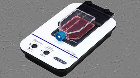
In this webinar, expert Joanna Hawryluk explores how the OLYMPUS Provi™ CM20 incubation monitoring system can help improve the health and stability of cell cultures through machine learning. With the aid of AI, the CM20 monitor automatically measures cell conditions using constant analysis parameters to provide quantitative data—all while your cultures remain safely in the incubator.

Hyperspectral and Brightfield Imaging Combined with Deep Learning Uncover Hidden Regularities of Colors and Patterns in Cells and Tissues
Experten
The Australian Research Council Centre of Excellence for Nanoscale Biophotonics draws on key advances of the 21st century, nanoscience, and photonics to help understand life at the molecular level. In this presentation, next-generation technologies developed in our Centre for probing, imaging, and interacting with the living systems will be discussed. These address the key challenges of ultrasensitive detection of key analytes in real complex environments and molecular complexity, and they support both novel therapies and diagnostics.

The Use of Multiplexing in Microscopy for Better Understanding the Skin Immune System in the Context of the Tissue
Experten
The skin is the first line of defense and the immune system’s biggest barrier for combating pathogens. Being able to accurately characterize and identify immune cell subtypes, tissues structures, and cell distribution in the skin under steady-state conditions provides a powerful tool for understanding the first immunological strategies and biological processes that occur in the presence of pathogens. In this webinar we will review technical aspects involved in the experimental process and explore how complementary imaging technologies might assist us to better understand the immune system.
The presentation is divided into three parts. First, an introduction of the Hugh Green Cytometry Centre will be presented and an overview of the histology and bioimaging technological platforms available. Second, the multiplexing methodology will be discussed, where several topics need to be considered for the design and development of a successful polychromatic panel for microscopy. Finally, preliminary results from a research project will be presented that constitutes part of a diploma program from The Royal Microscopical Society. The project focuses on the identification of immune cell types in the whole mount skin in relation to tissues structures (e.g., blood vessels and lymphatic network). It also centers on the immune cells’ distribution in the tissue as a first barrier of defense against pathogens.

Now You Have the Power to See More
Experten
The Olympus VS200 digital slide scanner has been very well received since its release in March 2020. With a reliable, flexible, and customizable design, the system has been adopted by various industries including research, geology, and many others. View this session to find out more and see examples of samples scanned using this popular addition to the Olympus product range.

Product Demo: SLIDEVIEW™ VS200 Research Slide Scanner
Experten
The Olympus SLIDEVIEW VS200 research slide scanner captures high-quality virtual slide images and enables advanced quantitative image analysis. Reliable virtual slide data can be acquired with as few as two clicks. Highly versatile, the SLIDEVIEW VS200 slide scanner supports five observation methods and a wide range of sample sizes for use in various applications. Its automatic slide loader accommodates many slide glasses, helping increase experiment efficiency.

Recent Advances in 3D Imaging and AI-Driven Data Analysis
Experten
This presentation will highlight various imaging techniques for 3D models, immunostaining with tissue clearing, and live imaging of organoids as well as AI-driven data analysis for high-content imaging and screening.


