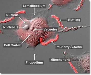
Found in virtually all eukaryotic cells, actin is a highly conserved globular protein that differs in amino acid sequence by less than 20 percent in species ranging from algae to humans. Actin is one of three major components of the cytoskeleton (along with tubulin and intermediate filaments) and is involved in a variety of important cellular functions, including muscle contraction, cell motility, division, organelle tranlation, signaling, and the establishment of "communication" junctions between cells. The confocal digital video presented above captures several rabbit kidney epithelial cells (RK-13 line) as they progress across the glass coverslip, extruding and contracting the leading edges of their lamellipodia, a process that mimics ocean waves crashing onto the beach. The actin in this specimen is labeled with mCherry fluorescent protein.
Video 1 - Run Time: 67 Seconds
Video 2 - Run Time: 58 Seconds
Video 3 - Run Time: 20 Seconds
Video 4 - Run Time: 67 Seconds
Video 5 - Run Time: 67 Seconds
Video 6 - Run Time: 58 Seconds
Video 7 - Run Time: 58 Seconds
Actin filaments are found throughout the cytoplasm of the cell but are concentrated just underneath the surface of the plasma membrane in the cortex. From the cortex, actin filaments spread out in all directions to lend support to the cell structure. Whereas microtubules direct intracellular traffic, actin filaments are responsible for the cell's overall movement in extra-cellular media. An actin filament is made of a 2-stranded helix, which may be several micrometers in length with a diameter of 5 to 9 nanometers. They are flexible structures that can be organized into bundles, networks, or three-dimensional gels. The confocal digital video presented above captures several rabbit kidney epithelial cells (RK-13 line) as they progress across the glass coverslip, extruding and contracting the leading edges of their lamellipodia, a process that mimics ocean waves crashing onto the beach. The actin in this specimen is labeled with mCherry fluorescent protein.

Among the many mechanisms involved in cell motility, actin filaments play a crucial role in cell movement during the extrusion of pseudopodia. A pseudopodium, or “false foot”, is a protrusion of continually assembling and disassembling actin filaments that extends and contracts to move the cell along. Myosin is involved with motility in the trailing end of the cell, causing it to contract by shuffling the cellular fluid to the front and into the pseudopodium, thus breaking the actin filament network. The pseudopodium then extrudes out from the cell body while the filament network rebuilds itself only to be contracted and broken again by the myosin. Many cells, including white blood cells, travel by extending pseudopodia, a phenonenon which is also known as crawling. The confocal digital video presented above captures several rabbit kidney epithelial cells (RK-13 line) as they progress across the glass coverslip, extruding and contracting the leading edges of their lamellipodia, a process that mimics ocean waves crashing onto the beach. The actin in this specimen is labeled with mCherry fluorescent protein.