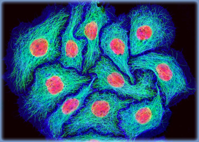
HeLa Cervical Carcinoma Cells Labeled with Alexa Fluor Dyes and TO-PRO-3
Immunofluorescence with mouse anti-alpha-tubulin was employed to visualize details of the microtubule network in a log phase monolayer culture of HeLa adenocarcinoma cells*. The secondary antibody (goat anti-mouse IgG) was conjugated to Alexa Fluor 488 and mixed with Alexa Fluor 546 conjugated to phalloidin to simultaneously image tubulin and the actin cytoskeleton. Nuclei were counterstained with TO-PRO-3, a carbocyanine monomer with long-wavelength red fluorescence.
*Although it became one of the most important cell lines in medical research, it’s imperative that we recognize Henrietta Lacks’ contribution to science happened without her consent. This injustice, while leading to key discoveries in immunology, infectious disease, and cancer, also raised important conversations about privacy, ethics, and consent in medicine.
To learn more about the life of Henrietta Lacks and her contribution to modern medicine, click here.
http://henriettalacksfoundation.org/
对不起,此内容在您的国家不适用。