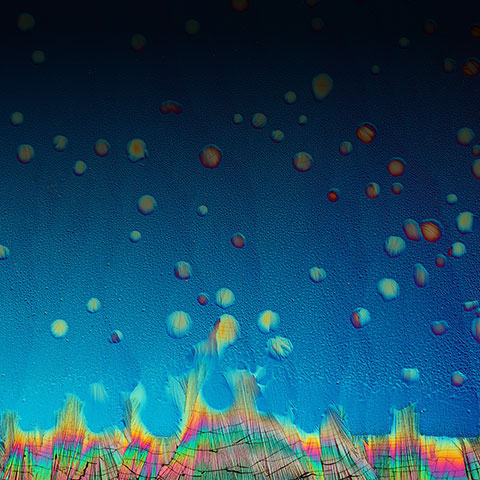
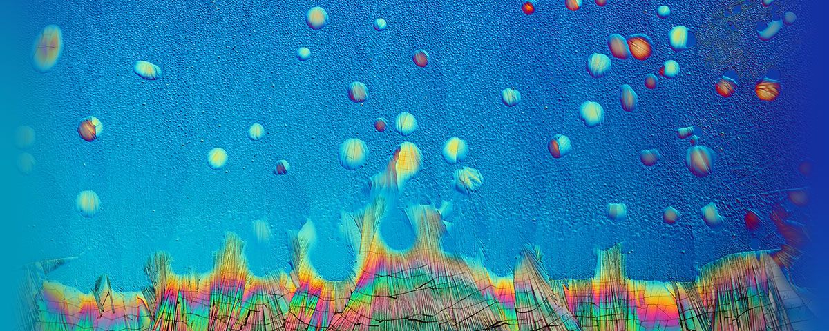
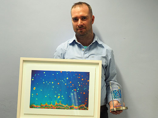
| Karl Gaff’s image of dopamine droplets on glass won second prize in the Olympus Image of the Year award 2018. Here he explains the optical effects and ingenious contrast methods that produce the spectacular colors from an otherwise translucent sample. |
|---|
Tell us about your pictureI call this picture ‘Ice Rain’. The image shows solidified microdroplets of the neurotransmitter dopamine on a glass substrate. I put a small quantity of dopamine on a glass slide and heated it so that when it melted it sizzled and splattered across the substrate forming small pools and tiny droplets. You can see the bulk crystal running along the bottom of the frame. Cracks in the crystal appear because of contraction as it cools. Regions that appear orange or magenta correspond to the thickest films while the silver and blue regions are the thinnest. The striking colors are produced by a combination of birefringence of the crystal structure and optical interference. For this to occur, the thickness of the film must be similar to the wavelength of the light. How did you create the image?I captured the image at a magnification of 100x using an Olympus BX51 microscope. The optical train of the microscope was configured for differential interference contrast (DIC). The dopamine film prepared on a glass substrate is translucent, but when placed in the DIC light path you can see the individual crystal grains and the dazzling interference colors they produce from their varying thicknesses and respective orientations. You can adjust the colors by translating the Nomarski prism, positioned above the objective, back and forth. The image was framed by rotating the camera and optimizing the detection parameters. How did you develop the idea for this picture?When I set out to capture photographs of crystals, I do not have a specific goal in mind. It is purely a research and discovery process in the sense that you experiment with different ratios of substances, heating and cooling temperatures as well as environmental conditions. It’s all about finding which permutations provide the most interesting and aesthetic formations. Of course, it’s never possible to reproduce those crystal structures exactly, so every slide is unique. It’s a matter of configuring and adjusting the components in the optical train together with composition to create an image that I am happy with. How did you get the idea to use a microscope as a tool for art?I was probably about eight years old when I received my first microscope. Even though it was a toy, I was mesmerized by what I could see. The microscope was incapable of having a camera adapted to it at the time. Of course, as I look back, it would have also been far from capable of producing the dazzling results that I am used to today. Over the years, I slowly upgraded with the dream of someday becoming a microscopist to effectively capture scientific yet beautiful artistic imagery using optical and electron microscopes. How long have you been combining microscopy and photography?It’s only in the past couple of years that I achieved my dream in gaining access to these cutting-edge research grade optical and electron microscopes that I now use to produce this unique photographic art. Since then, I have invested a lot of time in developing my knowledge and technique to make this imagery. I find it a very enjoyable and rewarding experience as I always see something new, develop my techniques and learn new things. What do you find most fascinating about microscopy?I am really interested in the physics of microscopes and how they work and the various configurations in which they can be used to probe the structure of materials. I am also fascinated by recent advances in super-resolution fluorescence microscopy, cryo-electron microscopy and X-ray diffraction. How they enable us to unravel the intricate architecture of the molecular nanomachines that exist in all cells, allowing us to discover the patterns of organization of biomolecules and how they come together to make up the organelles within cells. Where does this fascination stem from?From as far back as I can remember, I was curious and inquisitive about science. I suppose my fascination for microscopy and science comes from the first time I witnessed a drop of pond water magnified 100 times. An otherwise unremarkable drop of water to the naked eye, it contained a very large number of tiny machine-like animals going about their daily business, oblivious to the fact that they were being watched. How does microscopy help you in your work?The microscope allows me to explore samples collected from nature, to resolve their features in fine details and to enhance their contrast by using various configurations like darkfield, polarized light and DIC. By adapting electronic detectors, like a digital camera, I can take photographs of features of interest from both a scientific and an artistic perspective. What are you hoping to achieve inthe future?I hope to one day establish a microscopy lab specifically for creating these kinds of art works and to have my work used in university science text books. |
对不起,此内容在您的国家不适用。
对不起,此内容在您的国家不适用。


