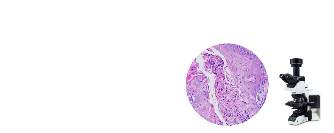
Designed for Laboratory Applications
The BX53 microscope combines a bright LED with suitable color temperature, automatic brightness adjustment, and ergonomic components so you can view samples in true-to-life colors while staying comfortable while you work.
Modular components enable you to expand as needed. You can upgrade your system for fluorescence, motorization, passive coding, multiple cameras, and optogenetics.
Fast and EfficientImproved Image QualityThe microscope is equipped with our advanced LED illuminator, equivalent to the brightness of a 100 W halogen lamp with a color temperature suitable for pathology and cytology. Simplify Your WorkflowWhen you switch objectives to change the magnification, the Light Intensity Manager automatically adjusts the brightness, helping save you time. | *Drag the arrow to compare brightness |
Pathological Research Applications
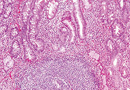 Tissue specimen observation | 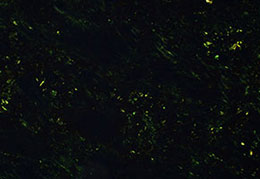 Amyloid/collagen observation | 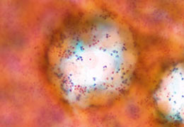 Cell specimen observation | 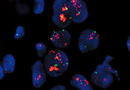 FISH observation |
Education and Conference
The BX53 microscope helps make training, education, and conference easy and efficient.
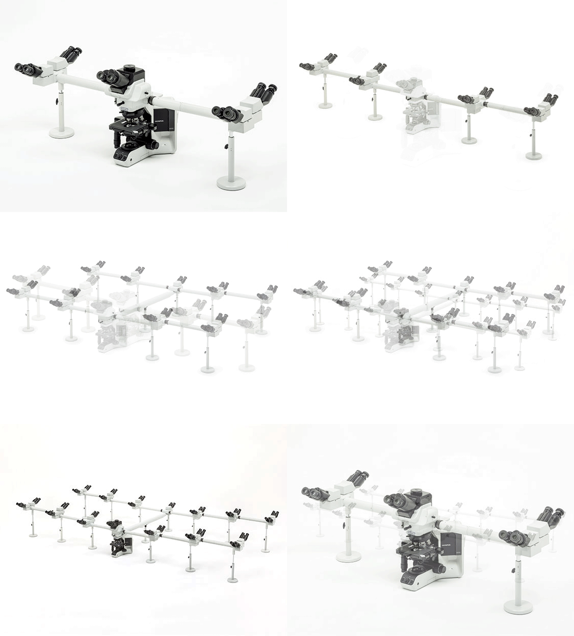
Add Up to 26 Observation HeadsMulti-head discussion systems are invaluable for lab training and education. With extremely small light loss and images that are oriented the same way at each observation head, everyone can observe the same image. Our multi-discussion observation (MDO) system can accommodate from 2 to 26 people. Powerful Digital ImagingAdd our full line of digital cameras to your system to share images with colleagues or for teaching. |
|---|
Bright Fluorescence Images
Expand your system to include multichannel fluorescence with up to 8 channels with no filter wheel.
The objectives' high transmission rates and fly eye illuminator provide bright images with even illumination.
Advanced Imaging with cellSens Software
Real-Time Panoramic ImagingCreate high-resolution, whole-slide images in real time simply by moving the controls of your manual stage with the Instant Multiple Image Alignment (MIA) function. |
Know Your Location on the SampleWhen imaging under high magnification, the position navigator enables you to see where you are on the overall sample slide. |
|---|
对不起,此内容在您的国家不适用。
对不起,此内容在您的国家不适用。

