Evident LogoOlympus Logo

咨询专家

The Use of Multiplexing in Microscopy for Better Understanding the Skin Immune System in the Context of the Tissue

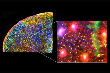
The skin is the first line of defense and the immune system’s biggest barrier for combating pathogens. Being able to accurately characterize and identify immune cell subtypes, tissues structures, and cell distribution in the skin under steady-state conditions provides a powerful tool for understanding the first immunological strategies and biological processes that occur in the presence of pathogens. In this webinar we will review technical aspects involved in the experimental process and explore how complementary imaging technologies might assist us to better understand the immune system.

The presentation is divided into three parts. First, an introduction of the Hugh Green Cytometry Centre will be presented and an overview of the histology and bioimaging technological platforms available. Second, the multiplexing methodology will be discussed, where several topics need to be considered for the design and development of a successful polychromatic panel for microscopy. Finally, preliminary results from a research project will be presented that constitutes part of a diploma program from The Royal Microscopical Society. The project focuses on the identification of immune cell types in the whole mount skin in relation to tissues structures (e.g., blood vessels and lymphatic network). It also centers on the immune cells’ distribution in the tissue as a first barrier of defense against pathogens.

Presenter: Alfonso J. Schmidt
Senior Staff Scientist, Malaghan Institute of Medical Research

Alfonso has a decade of experience working in a Shared Resource Lab (SRL) with a vast knowledge in histology, fluorescent microscopy, and image analysis. His work has been focused in maximizing the capabilities of the equipment available and in creating technical protocols and training modules for the scientific community. Currently, Alfonso oversees the Histology and Bioimaging Facility as part of the Hugh Green Cytometry Centre (HGCC) at the Malaghan Institute of Medical Research. Wellington, New Zealand.  

相关产品

激光扫描共聚焦显微镜

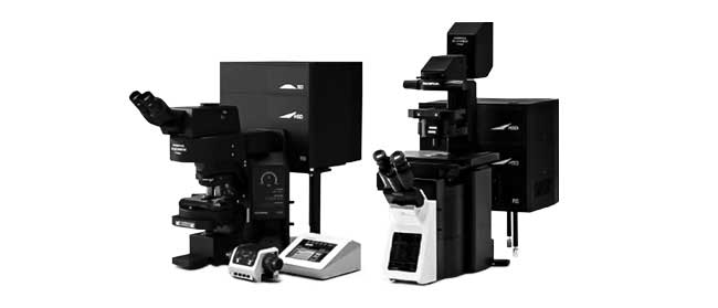
FV3000

  • 配有常规扫描单元的FV3000或配有常规/共振混合扫描单元的FV3000RS
  • 可在所有通道上进行高效、准确的全真光谱检测
  • 具有针对活细胞成像进行优化的高灵敏度和低光毒性
  • 倒置和正置镜架选项可适应各种应用和样品类型
研究级全玻片扫描系统

VS200

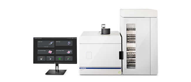
VS200研究级玻片扫查器提高了速度和效率,适用于需要定量分析大量样品的应用,包括脑、癌症、干细胞和药物发现研究。快速轻松地捕获高分辨率、高质量的玻片图像,并轻松地分析、共享和归档数据。VS200系统提供五种成像模式:明场、荧光、暗场、相衬和简单偏光。一次最多可加载210张载玻片进行自动扫描。

  • 2倍至100倍范围的高分辨率全玻片成像
  • 可顺应多种玻片尺寸和观察方法
  • 从明场到荧光多路传输的简化而又强大的工作流程
  • 从手动明场扫描到AI识别和全自动扫描,均可在软件和硬件中进行独特配置

The Use of Multiplexing in Microscopy for Better Understanding the Skin Immune System in the Context of the Tissue

The skin is the first line of defense and the immune system’s biggest barrier for combating pathogens. Being able to accurately characterize and identify immune cell subtypes, tissues structures, and cell distribution in the skin under steady-state conditions provides a powerful tool for understanding the first immunological strategies and biological processes that occur in the presence of pathogens. In this webinar we will review technical aspects involved in the experimental process and explore how complementary imaging technologies might assist us to better understand the immune system.

The presentation is divided into three parts. First, an introduction of the Hugh Green Cytometry Centre will be presented and an overview of the histology and bioimaging technological platforms available. Second, the multiplexing methodology will be discussed, where several topics need to be considered for the design and development of a successful polychromatic panel for microscopy. Finally, preliminary results from a research project will be presented that constitutes part of a diploma program from The Royal Microscopical Society. The project focuses on the identification of immune cell types in the whole mount skin in relation to tissues structures (e.g., blood vessels and lymphatic network). It also centers on the immune cells’ distribution in the tissue as a first barrier of defense against pathogens.

Presenter: Alfonso J. Schmidt
Senior Staff Scientist, Malaghan Institute of Medical Research

Alfonso has a decade of experience working in a Shared Resource Lab (SRL) with a vast knowledge in histology, fluorescent microscopy, and image analysis. His work has been focused in maximizing the capabilities of the equipment available and in creating technical protocols and training modules for the scientific community. Currently, Alfonso oversees the Histology and Bioimaging Facility as part of the Hugh Green Cytometry Centre (HGCC) at the Malaghan Institute of Medical Research. Wellington, New Zealand.  

相关产品

激光扫描共聚焦显微镜

FV3000

  • 配有常规扫描单元的FV3000或配有常规/共振混合扫描单元的FV3000RS
  • 可在所有通道上进行高效、准确的全真光谱检测
  • 具有针对活细胞成像进行优化的高灵敏度和低光毒性
  • 倒置和正置镜架选项可适应各种应用和样品类型
研究级全玻片扫描系统

VS200

VS200研究级玻片扫查器提高了速度和效率,适用于需要定量分析大量样品的应用,包括脑、癌症、干细胞和药物发现研究。快速轻松地捕获高分辨率、高质量的玻片图像,并轻松地分析、共享和归档数据。VS200系统提供五种成像模式:明场、荧光、暗场、相衬和简单偏光。一次最多可加载210张载玻片进行自动扫描。

  • 2倍至100倍范围的高分辨率全玻片成像
  • 可顺应多种玻片尺寸和观察方法
  • 从明场到荧光多路传输的简化而又强大的工作流程
  • 从手动明场扫描到AI识别和全自动扫描,均可在软件和硬件中进行独特配置
Experts
Alfonso J. Schmidt
Senior Staff Scientist
Malaghan Institute of Medical Research

Alfonso has a decade of experience working in a shared resource lab (SRL) with a vast knowledge in histology, fluorescent microscopy, and image analysis. His work has been focused in maximizing the capabilities of the equipment available and in creating technical protocols and training modules for the scientific community. Currently, Alfonso oversees the Histology and Bioimaging Facility as part of the Hugh Green Cytometry Centre (HGCC) at the Malaghan Institute of Medical Research in Wellington, New Zealand.

The Use of Multiplexing in Microscopy for Better Understanding the Skin Immune System in the Context of the Tissue2024年11月21日
Sorry, this page is not available in your country
Ask the Expert Sign-up

By clicking subscribe you are agreeing to our privacy policy which can be found here.

对不起,此内容在您的国家不适用。

Sorry, this page is not available in your country