The Olympus Museum: Microscopes
Key products in the history of Olympus
 1590 1590 | 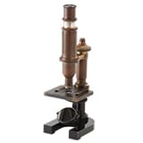 1920 ~ 1920 ~ | 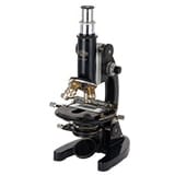 1931 ~ 1931 ~ | 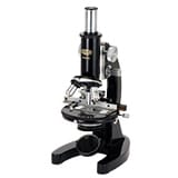 1946 ~ 1946 ~ | 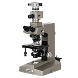 1972 ~ 1972 ~ |  1993 ~ 1993 ~ |  Inverted microscope Inverted microscope | 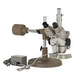 Stereo microscopes Stereo microscopes | 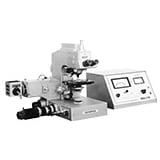 Quantification Quantification |  Photographic devices Photographic devices |
1590·1920 ~ The long road to locally produced microscopes
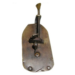
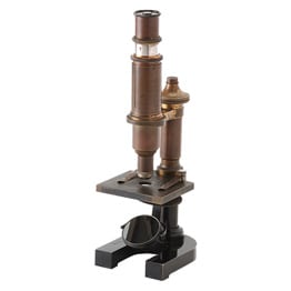
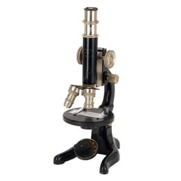
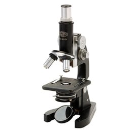
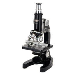
The history of microscopes begins with an invention by a Dutch father and son team of spectacle makers. Thereafter, further improvements were made in the UK and Germany. In Japan during the Meiji period, microscopes were produced and sold as magnifying glasses. However, they were inferior to European microscopes in terms of performance, and scientists that engaged in bacteriology research at that time had to rely on expensive imports.
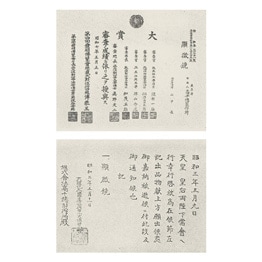
Takeshi Yamashita, the founder of Olympus, dreamed of somehow manufacturing microscopes in Japan. He established a company in 1919 and started working to fulfill his dream. This marked the beginning of a 13-year period of unswerving efforts by Yamashita.
1931 ~ Moving from monocular to binocular configurations
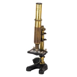
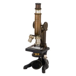
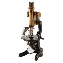
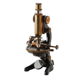
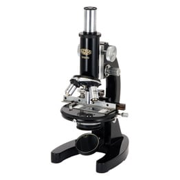
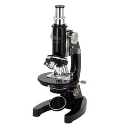
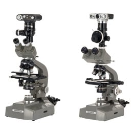
Olympus had built a full line-up of microscopes by the mid 1920s. From 1930, the company worked to make its products easier to use and to incorporate greater functionality, with the goal of integrating the following improved functionality and design elements:
- Mechanical stage to facilitate searches in the observation field
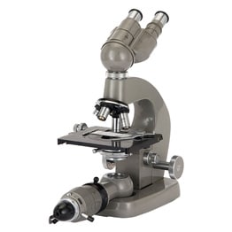
- Binocular heads that can be viewed using both eyes (making observations more comfortable)
- Improved optical performance through the development of apochromatic objective lenses
- Improved condenser performance
- Improved convenience when taking photographs
- Unified arm design
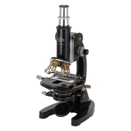 Fuji OCE: 1931 See details | 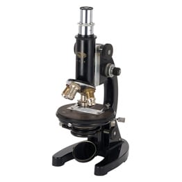 Kokka OCD: 1931 See details | 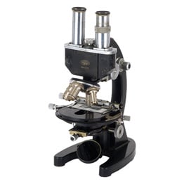 Mizuho LCE: 1935 See details | 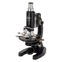 Homare UCE: 1935 See details | 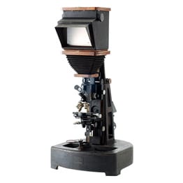 Super photo universal research microscope with a photographic system: 1938 See details |
1946 ~ Launch of the G Series
During the Second World War, the Olympus microscope and camera factory was relocated to an area of scenic beauty in Nagano prefecture to avoid damage from the war. The move was not a temporary relocation, but was planned as the construction of a new, regionally located factory for the long term.
In the postwar chaos, Olympus faced many problems. The company encountered various difficulties with "monozukuri" manufacturing at the Ina plant in Nagano prefecture that had taken over the role of the Hatagaya plant at the company's headquarters, which had been damaged during the war.
Olympus drew on its characteristic fortitude and spirit to resolve these problems and relied on its prewar models to achieve a new start at the Ina site. The strong performance by the microscope business today can be attributed to the ongoing strong determination of the company.
1972 ~ Diversifying microscopy needs
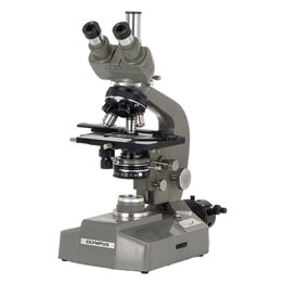
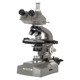
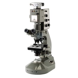
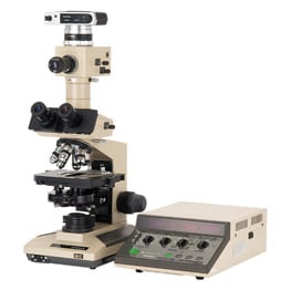
Microscopy needs diversified in line with developments in the sciences, engineering, and other fields. Olympus developed its microscopes to meet these demands by categorizing its portfolio according to their functional units. The company developed a main microscope body, which served as the platform, and incorporated it into the AH, BC, and CH series based on the application. Microscopes could now be created to suit specific objectives by combining various modules.
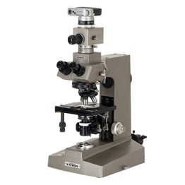 AH Series: 1972 See details | 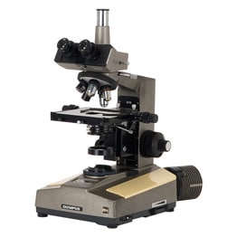 BH Series: 1974 See details | 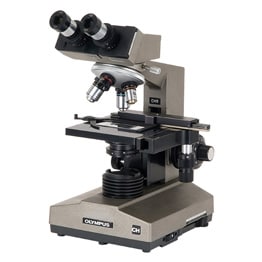 CH Series: 1976 See details | 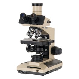 BH2 Series: 1980 See details | 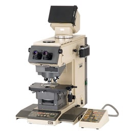 AH2 Series: 1983 See details |
1993 ~ Birth of the Y-shaped design
The objective lens determines the optical performance of a microscope. In a bid to improve lens performance, Olympus has always strived to improve its machining and assembly technologies. The company also pursued development with a focus on microscope design concepts, in a bid to respond to diverse demand from a wide range of fields.
The company's expertise in lens technologies and cutting-edge design concepts led to the development of the "UIS" optical concept that was used to create an innovative Y-shaped microscope design. This development highlighted the world-class ability of Olympus in this field.
 UIS objective lens: 1993 See details | 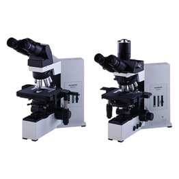 BX Series: 1993 See details | 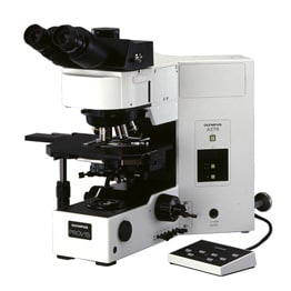 AX Series: 1994 See details | 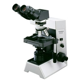 CX Series: 1997 See details |
Inverted microscopes: observing living cells
Microscopes come in two basic configurations: upright and inverted. Inverted microscopes are used to observe the specimen from below. They were first developed and used before the Second World War for research and analysis of metal materials such as iron and steel. With the advances made in biological research after the War, scientists started to use inverted microscopes for observing living cells.
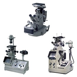 Inverted metallurgical microscope: 1954 See details | 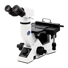 GX Series: 2001 See details | 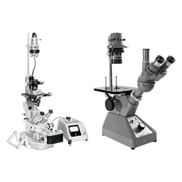 Inverted biological microscope: 1958 See details | 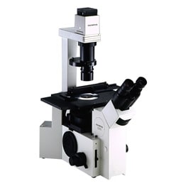 IX Series: 1994 See details |
Stereo microscopes: making 3D imaging possible
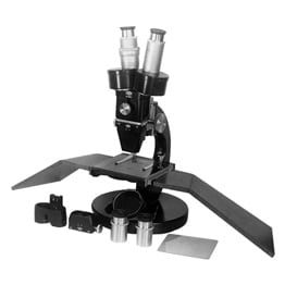
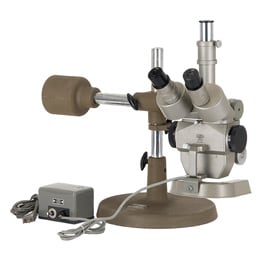
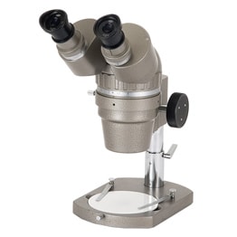
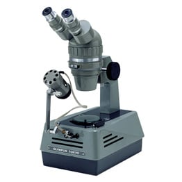
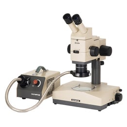
Humans, with two eyes, are able to see things in three dimensions. Stereo microscopes use this effect to make 3D imaging possible. Stereo microscopes are used for component assembly or precision part testing at factories because they allow visual confirmation of the object's surface irregularities or distances. Stereo microscopes have a long history, with first-generation models dating back to 1924. Due to demand, the models have evolved over the years to allow greater ease of use and better performance.
Quantification: expanding into various fields
Microscopes evolved from instruments for looking, examining, or recording into instruments for gauging or measuring.
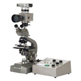
Evolving scientific needs drove the development of microscopes capable of quantification, for example through photometry or colorimetry. Such "color" data enabled significant advances in intercellular and genetic research. Moreover, microscope applications expanded into a number of spheres such as testing optical filters used in LCD TVs.
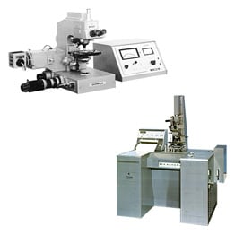 MMSP: 1971 See details | 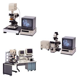 LSM Series: 1990, 1992 See details | 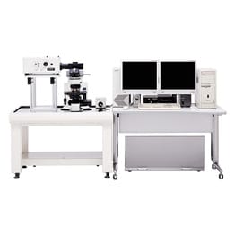 FV500/300: 1998 See details | 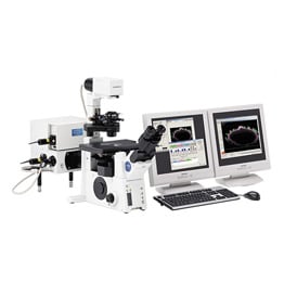 FV1000: 2004 See details |
Photographic devices: recording photographic images
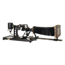
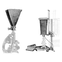
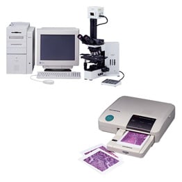
The arrival of digital cameras greatly simplified how microscope images and observations were recorded. Until then, microphotography was difficult and laborious for researchers, who squandered time learning the processes of selecting appropriate film, deciding on exposure time, and developing photographed images. To reduce the amount of time taken by researchers, microphotographic devices continued to evolve.
Sorry, this page is not
available in your country.

