Not Available in Your Country
Sorry, this page is not
available in your country.
Overview
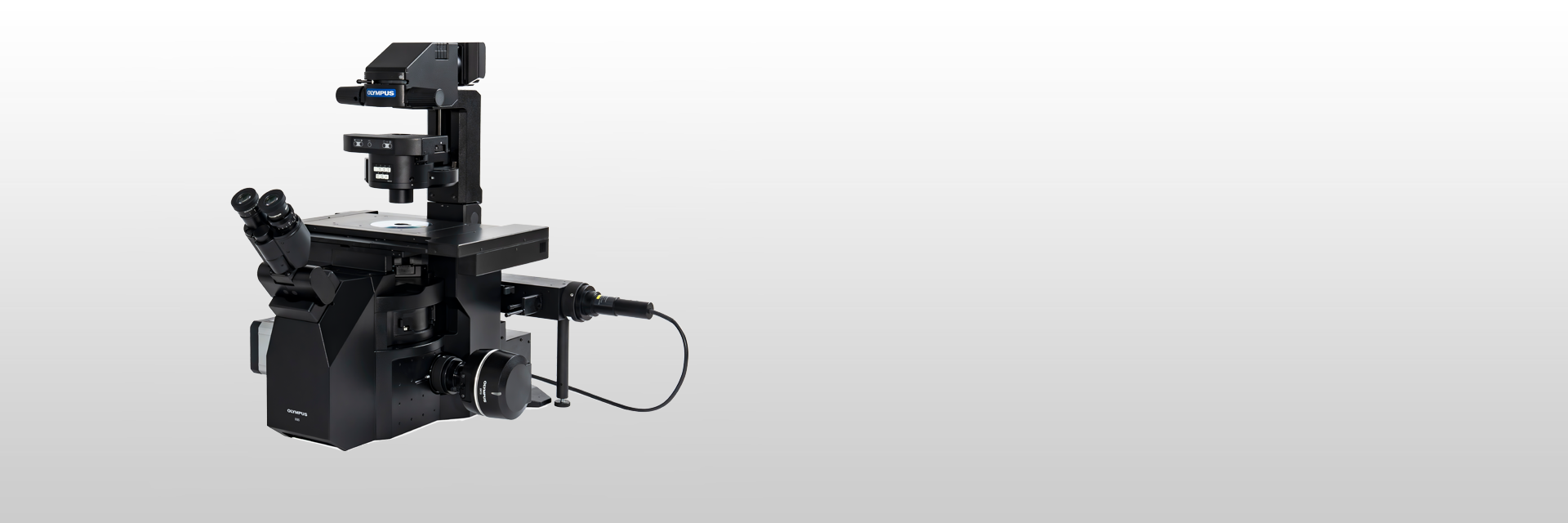
 | Clear, Fast, Accurate Live Cell ImagingThe IXplore™ IX85 Live inverted microscope system delivers precise live cell imaging, helps reduce photobleaching, and enhances cell viability for physiological experiments. And with an industry-leading 26.5mm field number (FN), the IXplore™ IX85 Live helps you acquire clear, accurate images faster than ever before. |
|---|
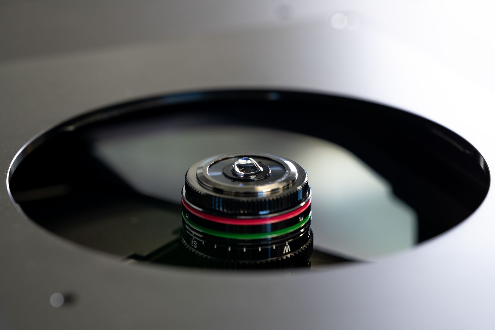
A New Standard in Image Depth and Objective UsabilityEvident’s silicone oil immersion objectives allow you to capture clearer images of living specimens during sophisticated timelapse experiments. These objectives help reduce the spherical aberration caused by refractive index mismatch and enable high-resolution observation deep inside living tissue. Our new multi-immersion objective (LUPLAPO25XS) introduces groundbreaking new immersion technology. This objective combines all of the advantages of our silicone oil immersion objective with new levels useability. With silicone gel pad technology, you can get the quality of silicone immersion oil with the useability of a dry objective.
| |
|---|---|
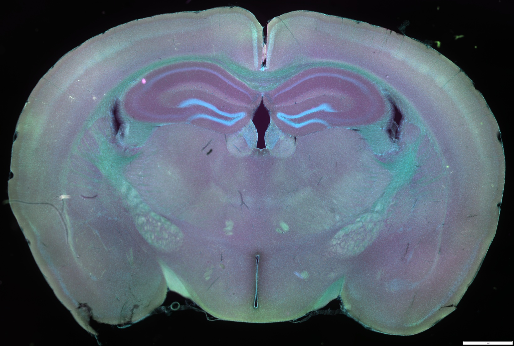
The new LUPLAPO25XS enhances workflows for organoids, 3D cell culture, well-plates, and a wide range of applications, with crystal-clear imaging and no compromise in usability. See deeper into your samples and reveal structures that were previously out of reach with a high NA and long working distance. | |
XYZ image comparison between Left: LUPLAPO25XS (Silicone gel) and Right: UPLXAPO20X (Dry)
|
Stable, Trustworthy Imaging OutputsLive cell imaging requires significant time and resources—it’s important that your lab has the right microscope system. The IXplore™ IX85 Live system offers enhanced rigidity and reduces the effects of vibration and temperature on your microscope. This also facilitates reliable timelapse imaging by helping maintain the desired focus position on the Z-axis. Pair the IXplore™ IX85 Live system with our TruFocus™ Z-drift compensator to capture cellular dynamics through high-precision, multipoint timelapse images that are never out of focus or misaligned. |
|---|
Carefully Maintaining Live SamplesLive cells require careful maintenance—we offer a variety of microscope-based incubation systems designed to meet your changing research needs. Box-type incubation systems* enable timelapse observations over several days by enclosing a portion of your microscope within the incubator. Shorter experiments can be completed with stage-top microscope CO2 incubation systems* that are fitted to your stage and can be easily removed when not in use by your team. Both incubation systems can be precisely controlled (temperature, humidity, and CO2 concentration) to maintain a constant environment surrounding your dish or well-plates. This maintains cell activity and significantly improves the reliability of your timelapse observations—ultimately providing you with better data. *Third-party products. | See how Jutta Bulkescher, Microscopy Specialist at the Center for Protein Research/Danish Stem Cell Center, University of Copenhagen, conducts a wide range of research at her facility and how incubation systems enables her to reliably perform stem cell analysis while maintaining cells under strict conditions. |
Cultured Cos 7 cell
| Close Monitoring of Cell Migration GrowthUse our cellSens Object Tracking and Count and Measure solutions to analyze the movement and division of live cells in timelapse or Z-stack image sets. Confluency Checker tools are a proven way for you to measure confluency on phase contrast images as well as fluorescence. |
|---|
Improve Experiment Efficiency with Advanced DeconvolutionWith our cellSens Dimension software, you can utilize live 2D deblurring for preview and acquisition to enable exceptional focusing on your thickest specimens. More advanced TruSight deconvolution is also available that uses an iterative algorithm to produce improved resolution, contrast, and dynamic range. To further improve experiment efficiency, you can define deconvolution processing as a macro function in the Graphical Experimental Manager (GEM). |  Esquerda: sem TruSight/Direita: com TruSight |
|---|
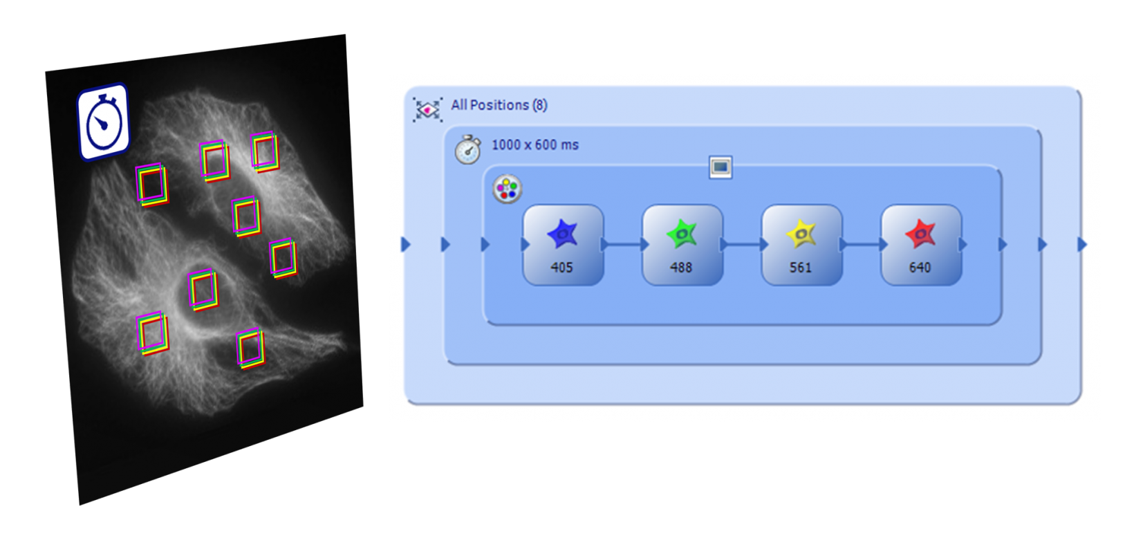
Fully Automated EfficiencyThe Graphical Experimental Manager (GEM) of cellSens Dimension software allows fully automated multidimensional observation (X, Y, Z, T, wavelength, and positions) and helps make experiment setup easier than ever. To further increase efficiency, you can also define macro functions, including deconvolution processing, in the GEM. |
|---|
IXplore™ IX85 Automated Inverted Microscope PlatformThe foundation of our IXplore IX85 Live system, the IXplore™ IX85 delivers the largest FN in the industry and an array of advanced end-to-end imaging features, allowing you to see and capture more than ever before while dramatically reducing acquisition times. Experience exceptional speed, clarity, and reliability with the IXplore IX85 microscope system. |
See how Evident Microscopes Have been used in live cell researchS. Wakayama, et al. Chemical labelling for visualizing native AMPA receptors in live neurons. Nature Communications (April 7, 2017). S. N. Cullati, et al. A bifurcated signaling cascade of NIMA-related kinases controls distinct kinesins in anaphase. The Journal of Cell Biology (June 19, 2017). L. Gheghiani, et al. PLK1 activation in late G2 sets up commitment to mitosis. Cell Reports (June 6, 2017). D. Nakane and T. Nishizaka, et al. Asymmetric distribution of type IV pili triggered by directional light in unicellular cyanobacteria. PNAS (June 5, 2017). T. A. Redchuk, et al. Near-infrared optogenetic pair for protein regulation and spectral multiplexing. Nature Chemical Biology (March 27, 2017). S. Barzilai, et al. Leukocytes breach endothelial barriers by insertion of nuclear lobes and disassembly of endothelial actin filaments. Cell Reports (January 17, 2017). J. Humphries, et al. Species-independent attraction to biofilms through electrical signaling. Cell (January 12, 2017). A. Prindle, et al. Ion channels enable electrical communication in bacterial communities. Nature (October 21, 2015). K. G. Harris, et al. RIP3 regulates autophagy and promotes coxsackievirus B3 infection of intestinal epithelial cells. Cell Host & Microbe (August 13, 2015). |
*1 Although it became one of the most important cell lines in medical research, it’s imperative that we recognize Henrietta Lacks’ contribution to science happened without her consent. This injustice, while leading to key discoveries in immunology, infectious disease, and cancer, also raised important conversations about privacy, ethics, and consent in medicine.
|
IXplore Microscopesfoi adicionado com sucesso aos seus favoritosMaximum Compare Limit of 5 ItemsPlease adjust your selection to be no more than 5 items to compare at once |
Need assistance? |
Specifications
| IX85P1ZF | IX85P2ZF | |||
|---|---|---|---|---|
| Microscope frame | Optical system | UIS2 optical system | ||
| Revolving nosepiece |
Motorized 6-position revolving nosepiece (DIC slider attachable),
One position for Automated Correction Collar Simple water proof structure | |||
| Focus |
Stroke: 10.5 mm
Minimum increment: 0.01 um, Maximum nosepiece movement speed: 3mm/s | |||
| Intermediate Magnification Changer |
3 positions (Coded)
1X / 1.6X / 2X | |||
| Light path selection |
Motorized 4 positions
Eyepiece 100%, left 100%, right 100%, eyepiece 50%/left 50% | |||
| Deck insert layer | 1 layer | 2 layers | ||
| Maximum port field number |
Left/Right side port: FN26.5, BI port: FN22
Deck right side port: FN18 |
Left/Right side port: FN18, BI port: FN22
Deck right side port: FN18 | ||
| Focus compensator |
TruFocus
Z drift compensator |
Offset method (Focus search, one-shot focus, continuous focus),
Class 1 laser product, laser wavelength: 830nm | ||
| Transmitted light illuminator |
Pillar tilt mechanism (30 ° inclination angle, with vibration reducing mechanism),
Condenser holder (with with 88 mm stroke, refocusing mechanism), Field iris diaphragm adjustable, 4 filter holders Light source: High power LED light source | |||
| Observation tube | Widefiled (FN22) |
• U-TBI90BK Wide field tilting binocular
• U-BI90 Wide field binocular • U-TR30-2/U-TR30NIR Wide field trinocular | ||
| Stage | Motorized stage |
• IX5-SSA: Stage stroke: X: 116mm x Y: 78mm, maximum stage movement speed: 40mm/s, Knob controller
• 3rd party motorized stage | ||
|
Mechanical stage with right handle
Mechanical stage with left handle |
Stage stroke: X: 116mm x Y: 78mm,
stage position locking function | |||
| Right handle stage | Stage stroke: X: 50mm x Y: 50mm | |||
| Gliding stage | Upper circular stage 360 ° rotatable, 20 mm (X/Y) travel | |||
| Plain stage | 232 mm (X) x 240 mm (Y) stage size, stage insert plate exchangeable (ø110 mm) | |||
| Condenser |
Motorized long working
distance condenser |
W.D. 27 mm, NA 0.55, motorized turret with 7 position slots for optical devices
(3 positions for ø30 mm and 4 positions for ø38 mm), motorized aperture and polarizer | ||
|
Long working distance
universal condenser | NA 0.55, W.D. 27 mm 5 positions for optical devices (3 positions for ø30 mm and 2 position for ø38 mm) | |||
| Ultra long working distance | NA 0.3, W.D. 73.3 mm, 4 positions for optical devices (for ø29 mm) | |||
| Fluorescence illuminator |
L-shape-fluorescence
illuminator | L-shaped design with exchangeable FS and AS modules, slider shutter and ND filter poket | ||
| Fluorescence mirror turret |
Motorized fluorescence
mirror turret | Motorized turret with 8 positions, built-in shutter, simple waterproof structurer | ||
| Fluorescence light source |
• U-LGPS: LED and LDP light source, Class 1 laser product
• 3rd party LED light source | |||
| Control unit (IX5-MCZ) | Nosepiece position, light path selection, filter turret position, FL shutter ON/OFF, DIA LED power, DIA LED ON/OFF, 4 customizable button | |||
| Control box (IX5-CBH) | PC interface | USB (Type-C), RS-232C | ||
| Operating enviornment |
• Indoor use
• Ambient temperature: 5 ºto 40 ºC (41 º to 104 ºF) • Maximum relative humidity: 80% for temperatures up to 31 ºC (88 ºF), decreasing linearly through 70% at 34 ºC (93 ºF), 60% at 37 º C (99 ºF), to 50% relative humidity at 40 ºC (104 ºF) • Supply voltage fluctuations: Not to exceed ±10% of the normal voltage | |||


