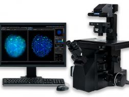
CKX41 Inverted Microscope
Discontinued Products
.jpg?rev=73BB)
This product has been discontinued, check out our current product CKX53
The CKX41 is an inverted microscope suitable for regular cell observation including GFP and other fluorescence applications. The high-angle tilting head is ideal for simple visual checks, while advanced Universal Infinity System 2 (UIS2) optics produce outstanding images. Olympus relief contrast increases visibility with non-glass cell culture vessels and improves productivity with its pre-centered slider. Post-acquisition analysis, documentation, and archiving can be achieved with digital cameras and software.
Not Available in Your Country
Sorry, this page is not
available in your country.
Características
Enhanced Cell Culture with Advanced UIS2 Optics
The CKX series make cell checking quicker and easier than ever before. Simple to operate, they require minimal optical adjustments and capture superb images with outstanding efficiency. A compact design allows installation inside cell culture clean benches and right beside the incubator, improving safety and saving time spent on transporting specimens for observation. Multiple observation methods are supported, and the range of applications has been further increased, with the CKX41 offering compatibility with numerous accessories. The CKX41 also features a tilting observation tube, allowing the user to use the system while standing, or a trinocular observation tube, compatible with a range of cameras.

UIS2 Optics Enable High Optical Performance
Through the preservation of image intensity, the UIS2 infinity-corrected optical system delivers improved resolution and contrast. Additionally, the maximum field of view has been extended to FN 22, allowing the usage of multiple upright observation tubes.
Cell Activity Status Observation with Enhanced Clarity
By simplifying the light path, UIS2 optics improve flatness for 10 to 15%, enabling clear, high contrast images that extend to the edge of the field of view.
Clarity at the Periphery with PHC-type Objectives

The PHC type objectives are minimally affected by the surface tension of the culture fluid, which compromises clarity at the image periphery. Multi-well observation is greatly improved from this feature. Combining this with the improved flatness from the UIS2 optics results in clear uniform observation at the cellular level.
Superior Optical Performance
High-clarity Relief Contrast Observation

Consistent directional contrast is maintained throughout all magnifications. Additionally, the slider is controlled by a simple lever and utilizes a common aperture between 20x and 40x, maximizing ease-of use and reducing time spent making optical adjustments.
Vertical Slider Installation Avoids Interference with Manipulators

Vertical slider installation prevents accidental touching of the manipulators while making optical adjustments.
Fluorescence Observation System
Effortless three channel fluorescence is accomplished with UIS2 filters (blue, green, UV) and objectives, while UV transmission is secured from an improved illuminator.


Quick, Adjustment-free Specimen Observation
Pre-centered Phase Contrast Slider for Quick, Adjustment-free Observation
Pre-centered phase contrast slider maximizes efficiency by eliminating the task of centering and exchanging ring slits when changing magnification. By minimizing the amount of optical adjustment, routine imaging can be completed easily.


Slim, Compact Design Takes Up Minimal Laboratory Space

Slim, compact design minimizes frame footprint, making installation effortless in any environment.
Tilting Binocular Tube for Flexible Observation

The binocular tube has a 30-60 degree tilting range, allowing for flexible observation and streamlined usage between incubator and microscope.
Simple Observation at the Clean Bench

Tilting binoculars greatly improves ergonomics when working at a clean bench.
Accessories
Stage Suitable for Hemocytometer and Other Plate Holders

Robust mechanical stage is compatible with hemocytometer inserts and other plate holders.
Glass Stage Insert Plate and Heat Plate

Glass stage and heat plate inserts are available to accommodate a variety of applications
Especificações
| Método de observação > Fluorescência (Excitação azul/verde) | ✓ | |
|---|---|---|
| Método de observação > Fluorescência (excitação ultravioleta) | ✓ | |
| Método de observação > Contraste de fase | ✓ | |
| Método de observação > Campo claro | ✓ | |
| Revólver porta-objetivas > Manual > Tipo padrão | Embutido, 4 posições | |
| Tubos de observação > Campo amplo (FN 22) > Tubo binocular com inclinação variável | ✓ | |
| Tubos de observação > Campo amplo (FN 22) > Trinocular | ✓ | |
| Platina > Manual > Platina simples | ✓ | |
| Condensador > Manual > Condensador com distância de trabalho ultra longa | NA 0,3/D.T. 72 mm (embutido) | |
| Dimensões (L × D × A) | 237.6 (W) x 515.5 (D) x 473.7 (H) mm | |
| Peso | 8,8 kg |
Componentes Relacionados
Color Cameras
Software

Fornecendo operações intuitivas e um fluxo de trabalho automático, a interface do usuário do software cellSens é personalizável e, assim, você controla o layout. Oferecido em uma série de pacotes, o software cellSens fornece uma variedade de recursos otimizados para as suas necessidades de formação de imagem específicas. Os recursos de Gerenciador de experimento gráfico e Navegador de poços facilitam a aquisição de imagem 5D. Obtenha uma resolução melhorada com a deconvolução TruSight™ e compartilhe suas imagens usando o modo de Conferência.
- Melhore a eficiência dos experimentos com a análise de segmentação com aprendizado profundo TruAI™, que fornece uma detecção de núcleos sem marcação e a contagem de células
- Plataforma de software de formação de imagem modular
- Interface do usuário orientada para a aplicação intuitiva
- Vasto conjunto de recursos, variando de uma simples imagem única a experimentos avançados multidimensionais em tempo real


