Judges 2013

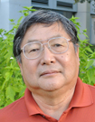
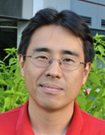
 | Dr. James Bear - Dr. Bear is a professor in the Department of Cell Biology and Physiology and a member of the Lineberger Comprehensive Cancer Center at the University of North Carolina (UNC), Chapel Hill. An HHMI Early Career Scientist, he also co-directs the UNC-Olympus Imaging Research Center at the university. In the laboratory he supervises, research focuses on the basic mechanisms of actin dynamics and cell motility, including the function of actin cytoskeleton regulators called coronins. In addition to having published dozens of papers throughout his career, he teaches, gives presentations, supervises graduate and post-graduate researchers and has served on numerous editorial boards. Among other honors, he was recognized in 2010 with the Hettleman Prize for Scholarly and Artistic achievement. |
 | Dr. Brian Matsumoto - Dr. Matsumoto is a well-known researcher, author and teacher and a recognized authority in the field of digital photomicrography. Adjunct Associate Professor at the University of California, Santa Barbara’s Neuroscience Research Institute and Department of Biology until his retirement in 2009, he was director of the university’s Integrated Microscopy Facility. The author of scores of papers and abstracts, and a reviewer for a number of academic journals, he has been a frequent presenter at microscope courses and educational forums. His images have appeared on the covers of numerous journals and his book Cell Biological Applications of Confocal Microscopy is considered a preeminent work on the subject. He and a colleague earned an honorable mention in the first Olympus BioScapes competition in 2004. |
 | Dr. Alison North - Senior Director of the Bio-Imaging Resource Center and Research Associate Professor at The Rockefeller University in New York City, Dr. North is a cell biologist with expertise in virtually all areas of fluorescence microscopy. Throughout her career, Dr. North has applied a variety of optical microscopy techniques to her research on cell-cell junctions and membrane-cytoskeletal interactions. Among the many advanced optical microscopy techniques she uses are 3D-SIM and STORM super-resolution techniques, laser scanning confocal, live-cell imaging, multiphoton, deconvolution, differential interference contrast, fluorescence recovery after photobleaching (FRAP), fluorescence resonance energy transfer (FRET), spinning disk confocal, laser microdissection, and a variety of techniques applied in digital image processing. Dr. North also has considerable experience with transmission and scanning electron microscopy. She says her favorite work activity of all is judging the Olympus BioScapes Competition. |
 | Dr. Lei Zhu - Associate Professor in the Department of Chemistry and Biochemistry at Florida State University, Tallahassee, Dr. Zhu heads a laboratory that studies a number of topics in organic and inorganic chemistry - in particular, the development of new fluorophores and new catalytic reactions, and the mechanistic characterizations of their functions. His research has resulted in dozens of publications in his field. Dr. Zhu was born in China and received his BS in chemistry from Peking University in 1997. After coming to the U.S., he worked with both James Canary of New York University and Eric Anslyn at University of Texas, Austin, before joining the faculty at FSU. |
Not Available in Your Country
Sorry, this page is not
available in your country.