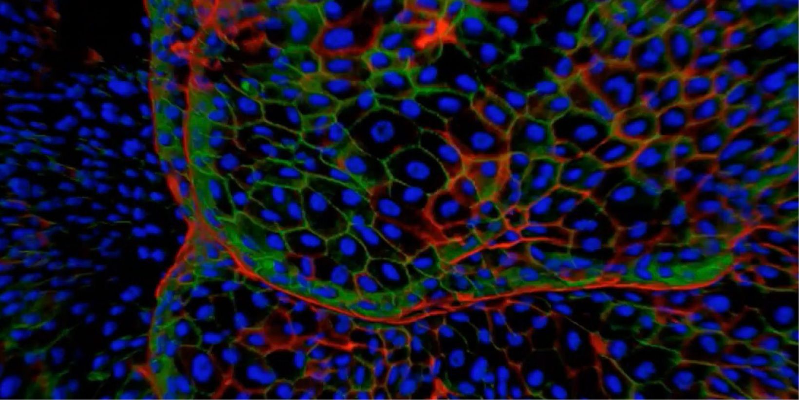
With rapid development in fluorescent proteins, synthetic fluorochromes, and digital imaging, advanced 3D imaging technologies are now available to investigators to provide critical insights into the fundamental nature of cellular and tissue functions. 3D and 4D imaging systems have become very common tools among biologists. However, there are several technical challenges and limitations in performing successful 3D and 4D imaging. Olympus has developed a wide range of 3D imaging microscopes to overcome these challenges and to satisfy the requirements of researchers across different disciplines.
In this talk, we will be taking you through the different imaging modalities that Olympus offers including widefield based fluorescence microscope like the Olympus IXplore and how in combination with image processing tools like Olympus TruSight can render usable 3D data sets.
However, more sophisticated 3D imaging systems are required for imaging samples such as organoids and spheroids. We will therefore illustrate the importance of laser-based systems that have been effectively used for deeper imaging into samples, like the Olympus FV3000 laser scanning confocal microscope, IXplore SPIN spinning disk confocal microscope and finally the FVMPE-RS multiphoton-excitation microscope. Along with these, we will stress the importance of high-quality objective lenses optimised for 3D imaging.

