Not Available in Your Country
Sorry, this page is not
available in your country.
Overview
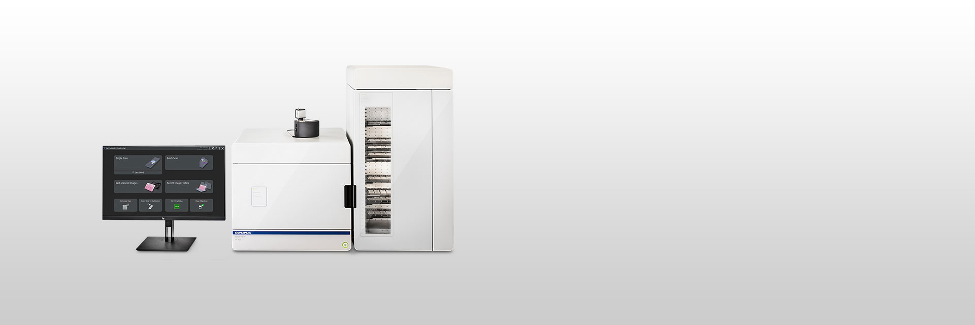 | The Power to See MoreEasily analyze, share, and archive your data with the SLIDEVIEW VS200 research slide scanner. Designed to capture high-resolution images of your slides for quantitative analysis, the system enables you to make the most of the information your slides have to offer. *This product is not available in some areas |
|---|
Outstanding Image Quality for QuantificationBetter Resolution and Flatness
|  |
|---|
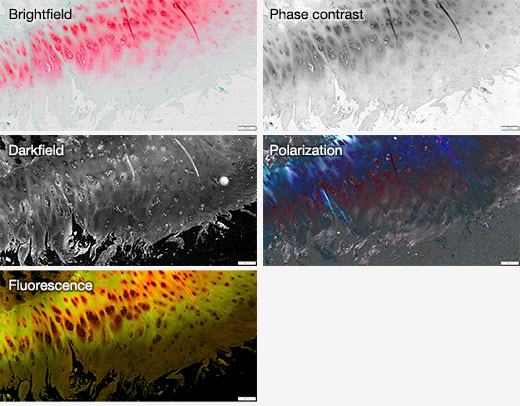
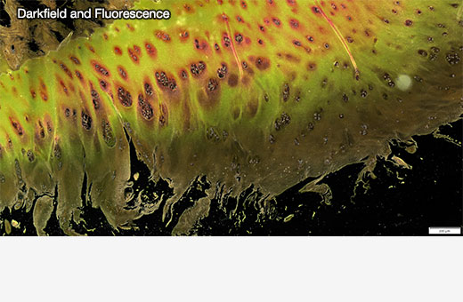
Flexible for Many ApplicationsSupports Multiple Hardware OptionsThe VS200 slide scanner is a highly flexible system that supports various slide sizes, different objectives, the SILA optical sectioning device, oil dispenser, cameras, loaders, and LED/laser light sources with multiple filter sets. Learn more about the SILA optical scanning device Supports Multiple Observation MethodsThe VS200 supports brightfield, darkfield, polarization, phase contrast, and fluorescence, with the possibility to scan a sample using multiple techniques at the same time. Flexible Software SolutionsThe VS200 software supports offline analysis, deep learning, 3D deconvolution, and batch conversion to standard file formats. It also supports the NIS-SQL database for data management and the DICOM format for easy integration into a lab information management system (LIMS).
|
Achieve More in Less Time
| Related Videos |
|---|
Simplified and Powerful Workflow
|
|---|
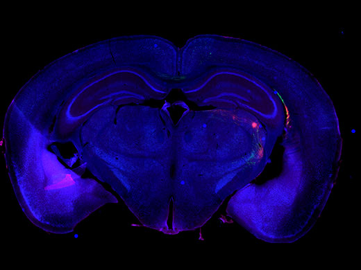
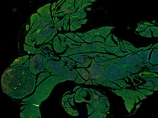
Reliable Data for Many ApplicationsThe flexibility of five imaging modes and outstanding Olympus X Line objectives make the VS200 slide scanner a solution for many applications.
|
Testimonials |
Michel Meijer
| “Within the slide scanning facility of the pathology department there was a high demand for a whole slide scanning microscope with multiple methods of illumination (brightfield, polarization, and fluorescence) and a diverse range of objectives (air and oil). The SLIDEVIEW VS200 scanner fits our needs as a day-to-day whole slide scanning system for research purposes without compromising on image quality. | |
Need assistance? |
The VS200 is not for clinical diagnostic use. |
Applied Technologies
High-Resolution Scanning
| Related Videos |
|---|
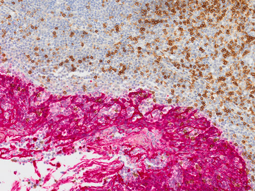
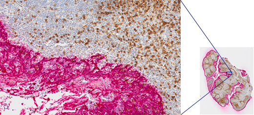
Tonsil CD3 (rm), ImmPRESS Reagent (HRP) Anti-Mouse IgG Immpact DAB (brown), AE1/AE3(m) ImmPRESS (AP) (HRP) Anti-Rabbit IgG Immpact Vector Red (red). Counterstained with Hematoxylin QS (blue).
| Accurate Focus at All Magnifications
|
|---|
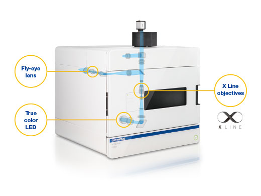
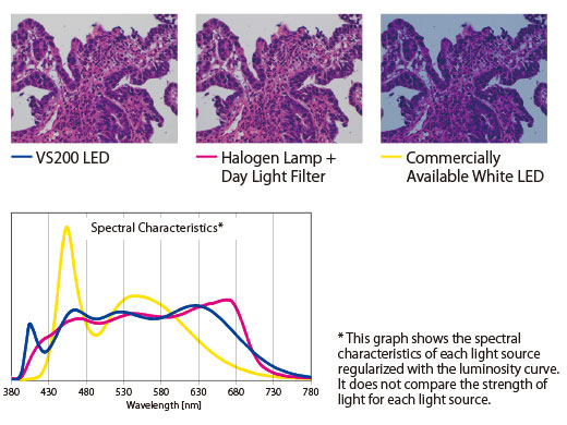
Bright LED with Accurate Color ReproductionThe system’s true color LED for transmitted illumination has the same spectral characteristics and power as a halogen lamp, so purple, cyan, and pink stains are correctly represented, imaged, and rendered. |
|
|---|
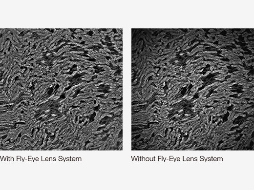 | Uniform Fluorescence IlluminationThe fluorescence illuminator with its fly-eye lens uniformly distributes light across the entire field of view for bright, evenly illuminated images. |
|---|
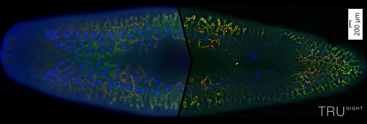
High-Contrast Images without BlurThe SILA optical sectioning device quickly delivers blur-free scans of thick samples, so you can achieve widefield and section imaging with a single system. Based on Hi-Lo microscopy, the SILA (speckle illumination acquisition) optical sectioning device produces high-contrast images free from out of focus blur. Adding the SILA hardware to a VS200 makes it especially well suited for imaging thicker samples that either normal or TruSight imaging cannot resolve effectively. The SILA device delivers real-time, blur-free scans of thick samples, so you can achieve widefield and section imaging with a single system. | Widefield (left), Widefield + TruSight Deblurring (middle) and SILA (right) images of a whole planarian flatworm Schmidtea mediterranea in 20x, showing the intestines. Blue: DAPI. Green: inner intestine cells; Red: outer intestine cells. Samples provided by Amrutha Palavalli, Department for Tissue Dynamics and Regeneration, Max Planck Institute for Multidisciplinary Sciences, Goettingen, Germany. |
Related Videos |
Online Fluorescence DeblurringTruSight Live deblurring reduces diffused light from above and below the focal plane of a thick sample. The image data is then recalculated using a special 2D deconvolution algorithm, making the images sharper and clearer. |
Without TruSight Live (left) vs. with TruSight Live applied to all three channels (DAPI, FITC, and CY3) (right). S. mediterranea stained with double fluorescent (red and green) in situ hybridization, counterstained with DAPI, and scanned at 10X magnification. Sample provided by Miquel Vila-Farré, Max Planck Institute of Molecular Cell Biology and Genetics. |
|---|
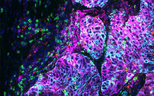
Lung tissue imaged on a VS200 at 20X stained with an Ultivue PD-L1 kit multiplex kit; Dapi: Nuclear Counterstain, FITC: CD8, TRITC: CD68, Cy5: PD-L1, Cy7: panCK.
| Multiplex Scan ModeWhen tissue samples are limited, it’s important to get the most possible data from each tissue section. Multiplexing mode aligns multiple fluorescence channels with a reference image, enabling greater understanding of co-expression and spatial composition of multiple targets within a sample. |
|---|
The Power of TruAI Deep-Learning Technology
|
Left: Cy3 fluorescence marked pancreatic islets. Pancreatic islets are stained (red) while erythrocytes are autofluorescencing.
Learn more about the application: Segmentation and Analysis of Pancreatic Islets |
Advanced Automation for More Efficient Image AcquisitionOptimized Slide Scanning Workflow
| Related Videos |
Workflow-Based Solution, Including Pretrained Neural Networks
Powerful Image Analysis with AI-Based SegmentationTruAI technology offers instance segmentation analysis to simplify workflows and rapidly deliver more accurate results. The deep neural network’s performance is superior to traditional segmentation techniques, and you can develop your own neural network libraries for different applications. Once trained, the neural network can be applied with a single click and shared with collaborators. |
.jpg?rev=22E1) .jpg?rev=AF00) | Higher ProductivityMultibatch Processing
Hot-SwapHot-swap functionality enables you to exchange an already scanned slide tray with a new tray during batch processing so that you can add new slides before the system has finished scanning. This function minimizes the amount of time the system is idle. |
|---|
Need assistance? |
Data Management
Comprehensive Image and Data Management
|  | |||||||||||||||||||||
|---|---|---|---|---|---|---|---|---|---|---|---|---|---|---|---|---|---|---|---|---|---|---|
The VSI file format supported by VS200 systems was developed by EVIDENT to meet the specialized requirements of advanced microscopy imaging. A number of projects and companies are already supporting the VSI format in their applications, and more are on the way.
| ||||||||||||||||||||||
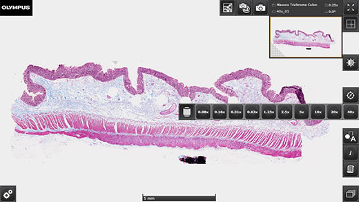
Colon stained with Masson's Trichrome. | Remote Access with Free OlyVIA Virtual Slide Viewer
|
|---|
Need assistance? |
Specifications
| VS200 Single Tray | VS200 Multiple Tray Loader | ||
|---|---|---|---|
| Intended Specimen | Observable Specimen | Glass slide with cover glass | |
|
Size of Glass Slide
(W × L × H) |
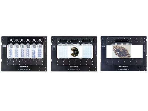
Standard slide tray: 25 mm–26.5 mm × 75 mm–76.5 mm × 0.9 mm–1.2 mm (1 in. × 3 in. × 0.05 in.) (6 slides)
Optional trays 1) 51 mm–53 mm × 75 mm–76.5 mm × 0.9 mm–1.2 mm (2 in. × 3 in. × 0.05 in.) (3 slides) 2) 100 mm–102 mm × 75 mm–76.5 mm × 0.9 mm–1.2 mm (4 in. × 3 in. × 0.05 in.) (1 slide) 3) 126 mm–128 mm × 75 mm–76.5 mm × 1.1 mm–1.4 mm (5 in. × 3 in. × 0.06 in.) (1 slide) | ||
| Cover Glass Thickness | 0.12–0.17 mm | ||
| Observation Methods | Brightfield, reflected brightfield (optional*1), darkfield, phase contrast (optional*2), simple polarization (optional*3), fluorescence (optional), fluorescence optical sectioning with speckle illumination (optional SILA module) | ||
| Optical Frame | Illuminator | Built-in Köhler illumination for transmitted light; high intensity and high color rendering LED (up to 50,000 hours) | |
| Objectives |
Compatible objectives 2x, 4x*4, 10x*5, 20x, 40x*5, 60x*5, and 100x*5 6-position motorized nosepiece (incl. selected oil immersion, silicon oil immersion, and phase contrast objectives)
Optional automatic oil dispenser | ||
| Motorized Stage | XY stage with automatic control | ||
| Focusing | Motorized focusing with automatic control | ||
| Color Camera | Integrated 2/3 inch CMOS, 3.45 μm × 3.45 μm pixel size, high sensitivity, high resolution | ||
| Scan Unit | Capacity | 1 slide tray, 6 slides maximum; upgradable to a multiple tray loader model | Up to 35 slide trays, 210 slides maximum |
|
Pixel Resolution
(Color Camera) |
UPLXAPO20X (NA 0.8): 0.274 μm/pixel
Options: UPLXAPO4X (NA 0.16): 1.37 μm/pixel UPLXAPO10X (NA 0.4): 0.548 μm/pixel UPLXAPO40X (NA 0.95): 0.137 μm/pixel UPLXAPO40XO (NA 1.4): 0.137 μm/pixel UPLXAPO60XO (NA 1.42): 0.091 μm/pixel UPLXAPO100XO (NA 1.45): 0.055 μm/pixel | ||
| Scan Time |
Brightfield: approx. 1.5 minutes (20x objective, scan area 15 mm × 15 mm)
| ||
| Software | Automatic sample detection (generic and TruAI deep learning), automatic barcode reading, automatic focus mapping, automatic scanning, automatic stitching, pause and resume scanning, Z-stack imaging, extended focus imaging (EFI), image format: vsi, JPEG, TIFF, DICOM, synchronized multi-image display, stepless zooming, zooming while scanning, annotations, screen capture, slide loader control (multiple tray loader only) | ||
|
Fluorescence
(optional) | Fluorescence Components |
UPLFLN4X objective, illuminator with fly-eye lens, motorized mirror turret, motorized filter wheel
Widefield light source options: U-LGPS, Excelitas X-Cite XYLIS, X-Cite TURBO, X-Cite Novem SILA: 4-line laser combiner (405 nm, 488 nm, 561 nm, and 638 nm) and scrambler unit | |
| Monochrome Camera |
Options:
VS-304M, 1-inch CMOS, 3.45 μm × 3.45 μm pixel size HAMAMATSU ORCA Flash4.0 V3 HAMAMATSU ORCA Fusion HAMAMATSU ORCA Fusion BT | ||
|
Solutions for Scanner Software
(optional) | Solution License |
Batch image format converter
DICOM converter Fluorescence SILA acquisition | |
|
Desktop Software
(optional, separate solution for analysis) | Solution License |
Batch image format converter
DICOM converter Detection and analysis Deep learning 3D deconvolution | |
| Environment | Weight |
Optical frame: 69 kg (152.1 lb)
1 slide tray: 0.6 kg (1.3 lb) |
Optical frame and multiple tray loader: 142 kg (313 lb)
35 slide trays: 21 kg (46.3 lb) |
|
Fluorescence: 8 kg (17.6 lb)
PC and monitor: 16 kg (35.3 lb) Camera cover (optional): 9 kg (19.8 lb) | |||
| Operating Environment |
Temperature: 15–28 °C (59–82.4 °F) (including other devices)
Humidity: up to 80% (31 °C (87.8 °F)) | ||
| Power Supply*5 |
Input: AC 100–240 V, 50/60 Hz, 4 A
Output: DC 24 V, 9.2 A | ||
| Power Consumption |
Input: 100–240 V AC; 50/60 Hz; 4 A
221 W | ||
*1 Optional phase contrast objectives are required.
|






![VS200 [Instructions for use]](https://static2.olympus-lifescience.com/modules/imageresizer/ceb/827/e0120c2836/112x84p67x50.jpg)


