Not Available in Your Country
Sorry, this page is not
available in your country.
Overview
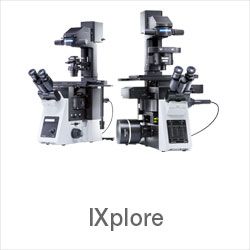
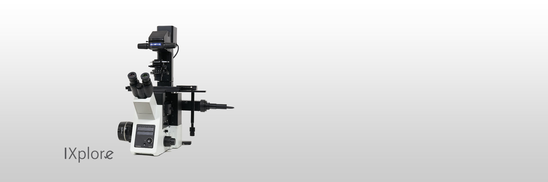 | High-Quality ImagingDesigned for simple multicolor fluorescence imaging and routine experiments, the IXplore™ Standard microscope system is easy to operate and capable of producing excellent, publication-quality images. A range of encoded unit options makes it easy to get accurate and reproducible results. |
|---|
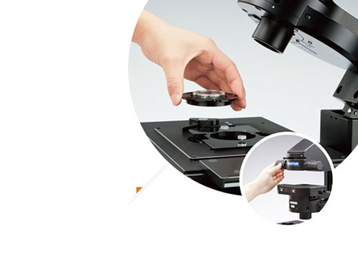 | Accuracy and RepeatabilitySmooth Tracking at High MagnificationThe IX3-SVR manual stage features a smooth positioning system that enables cells to be easily tracked, even at high magnifications.
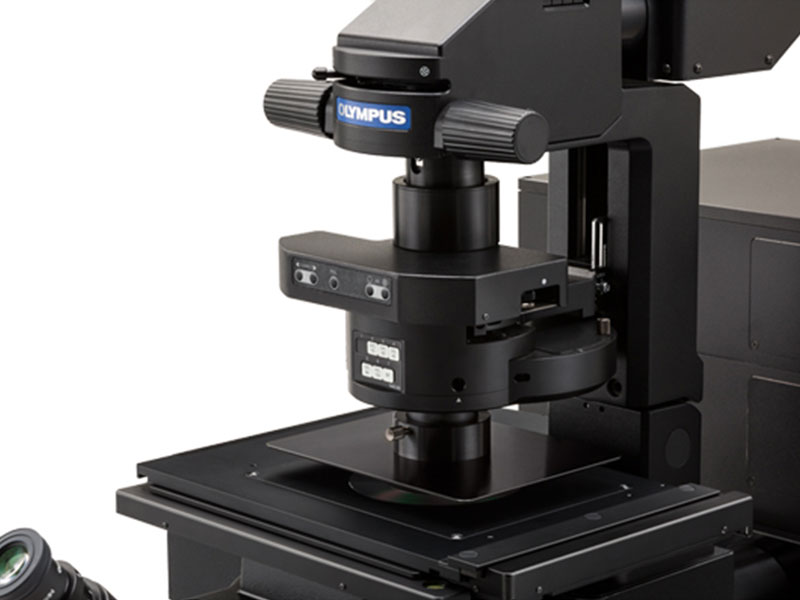
Easy Köehler IlluminationThe condenser can be moved and easily reset to Köhler illumination using the conveniently located condenser lock and control knobs. |
|---|
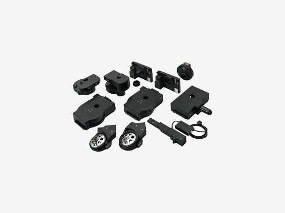
Encoded Units (Optional Peripherals)A Cost-Effective Way to Upgrade to a Smart MicroscopeA wide range of optional units are available for upgrades, including:
|  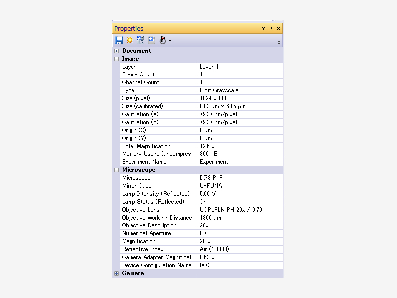 |
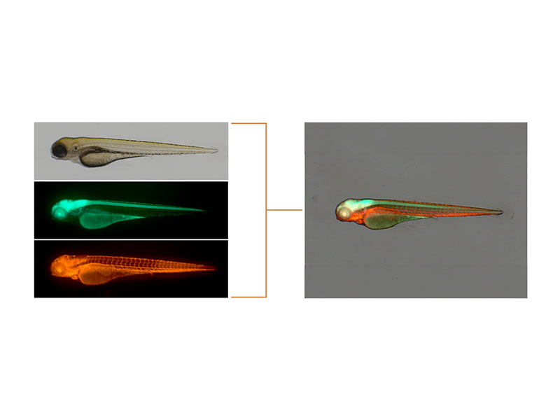  | Ease of UseOne Camera for Multiple ApplicationsThe DP75 digital microscope camera is a high-performance, multi-application imaging tool that makes it easy to capture high-resolution brightfield or fluorescence images using a single camera. High Contrast under Bright ConditionsThe unit is designed specifically for fluorescence observation. It efficiently blocks out room light, enhances the contrast of fluorescence, and enables clear fluorescence observation under bright conditions. Intuitive Operation with cellSens™ Imaging SoftwareOur cellSens software is easy to use, powerful, and flexible. Featuring a modular design, the software can be tailored to your budget and imaging applications. This enables the software to grow and adapt to meet evolving research needs. |
|---|
Simplify Your WorkflowObjectives for Observation Using Plastic VesselsLUCPLFLN series objectives, and in particular the UCPLFLN20XPH (NA 0.7), are well-suited for observation using plastic dishes. The objectives enable high-resolution observation of the cell proliferation process and deliver improved contrast across a wide area. This gives you the flexibility to image through plastic-bottom dishes in addition to glass. *Image: iPS-cell expressing Nanog reporter (GFP) Image data courtesy of: Tomonobu Watanabe, Ph.D. Laboratory for Comprehensive Bioimaging, RIKEN Quantitative Biology Center | .jpg?rev=79D5) .jpg?rev=44DA) 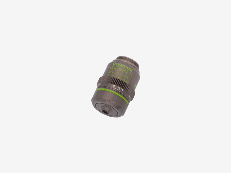 |
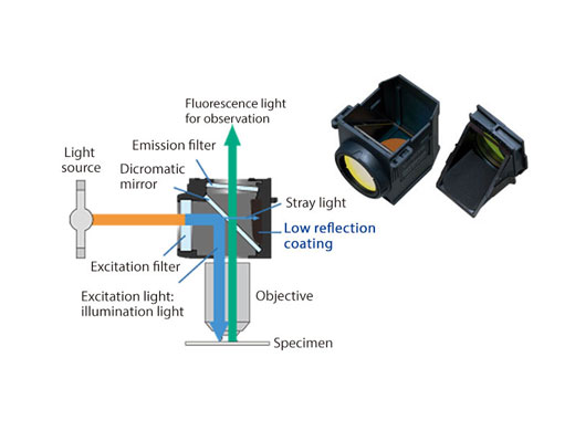 | High-Quality ImagingHigh Signal-to-Noise Fluorescence Mirror Units for Efficient Signal DetectionAll fluorescence mirror units feature filters treated with a specially developed coating that absorbs more than 99% of stray light. This reduction in reflections and high transmittance of the mirror units helps provide fluorescence images with a high signal-to-noise ratio. |
|---|
Need assistance? |
Specifications
| Microscope Frame | IX73P2F | |
|---|---|---|
| Observation Method > Fluorescence (Blue/Green Excitation) | ✓ | |
| Observation Method > Fluorescence (Ultraviolet Excitation) | ✓ | |
| Observation Method > Differential Interference Contrast (DIC) | ✓ | |
| Observation Method > Phase Contrast | ✓ | |
| Observation Method > Brightfield | ✓ | |
| Revolving Nosepiece > Motorized (6 position) | ✓ | |
| Revolving Nosepiece > Manual > Coded (6 position) | ✓ | |
| Observation Tubes > Widefield (FN 22) > Tilting Binocular | ✓ | |
| Illuminator > Transmitted Köhler Illuminator > LED Lamp | ✓ | |
| Illuminator > Transmitted Köhler Illuminator > 100 W Halogen Lamp | ✓ | |
| Illuminator > Fluorescence Illuminator > 100 W Mercury Lamp | ✓ | |
| Illuminator > Fluorescence Illuminator > Light Guide Illumination | ✓ | |
| Fluorescence Mirror Turret > Motorized (8 position) | ✓ | |
| Fluorescence Mirror Turret > Manual > Coded (8 position) | ✓ | |
| Stage > Motorized | Contact your local sales representative to hear about motorized stage options | |
| Stage > Mechanical > IX3-SVR Mechanical Stage with Right Handle |
| |
| Stage > Mechanical > IX3-SVL Mechanical Stage with Left Short Handle |
| |
| Condenser > Motorized > Universal Condenser | W.D. 27 mm, NA 0.55, motorized aperture and polarizer | |
| Condenser > Manual > Universal Condenser | NA 0.55/ W.D. 27 mm | |
| Condenser > Manual > Ultra-Long Working Distance Condenser | NA 0.3/ W.D. 73.3 mm | |
| Confocal Scanner | - | |
| Super Resolution Processing | - | |
| Accessories | - | |
| Dimensions (W × D × H) | 323 (W) x 475 (D) x 721 (H) mm (IX73 microscope frame) | |
| Weight | Approx. 47kg (IX73P2F) |


