Image Segentation
Fast and Efficient
- High-precision detection and segmentation based on deep-learning technology delivers efficient and reliable analysis results
- Optimal for cell counting and geometric measurements, like area or shape, without phototoxicity
- Less than 1-second processing time per position
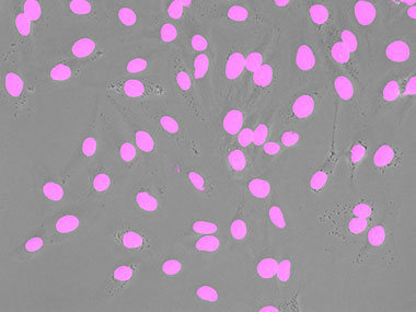
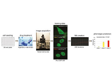
An example showing the detection of nuclei. A schematic showing the application (inference) of the trained neural network. |
Next-Generation Image Analysis Based on Deep Learning
Experiments often require data from microscope images. For accurate image analysis, segmentation—especially thresholds based on intensity values or color—is used to extract the analysis targets from the image. But this can be time-consuming and affect the sample condition.
TruAI technology’s next-generation image analysis helps solve these challenges.
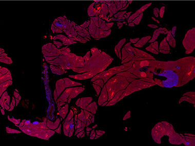
| Prediction of mitotic cells using TruAI (green). | Prediction of glomeruli positions on a mouse kidney section using TruAI (blue). | Green: You can see that the detection accuracy is low due to the unevenness of the GFP label.
Blue: Detecting the nuclei with high accuracy despite scratches and dust on the vessel. |
Image EnhancementThe neural network can learn the features of noise in advance, making it possible to construct images with a high signal-to-noise ratio, even if the signals are weak. Noisy images with very weak fluorescence intensity also make object recognition for segmentation difficult. It’s also important to minimize image fading and acquire the images as quickly as possible. |
Save Time and Effort with Live AIObserve as the live inference results of the trained neural network are revealed in real time. Knowing the inference results before you begin image acquisition increases the efficiency of your experiments. | Related VideosLive detection of different phases in the cell cycle with HeLa cell culture* |
*Although it became one of the most important cell lines in medical research, it’s imperative that we recognize Henrietta Lacks’ contribution to science happened without her consent. This injustice, while leading to key discoveries in immunology, infectious disease, and cancer, also raised important conversations about privacy, ethics, and consent in medicine.
To learn more about the life of Henrietta Lacks and her contribution to modern medicine, click here.
http://henriettalacksfoundation.org/
Macro to Micro ImagingThe macro to micro function enables you to acquire an overview image using low-magnification objectives, such as 4x, and then detect the sample area and acquire images of it at high magnification. By using TruAI, this process happens automatically, making your imaging faster and more efficient when using glass slides or dishes with multiple tissue sections. |
|
 | “The pretrained nuclei recognition is absolutely stunning and now makes it possible to easily analyze very heterogenous samples without compromising on any cell fractions. Especially in high cell density areas, TruAI-based separation is clearly superior to intensity or edge detection, both speed- and performance-wise.” Robert Strauss |
Learn More
Perform Accurate and Efficient Microscopy Image Analysis Using TruAI Technology with Deep Learning | Predicting Multi-Class Nuclei Phenotypes for Drug Testing Using Deep Learning | Rapid Automated Detection and Segmentation of Glomeruli Using Self-Learning AI Technology | Accelerating and Optimizing the Segmentation and Analysis of Pancreatic Islets with the VS200 Research Slide Scanner and TruAI Deep-Learning Solution |
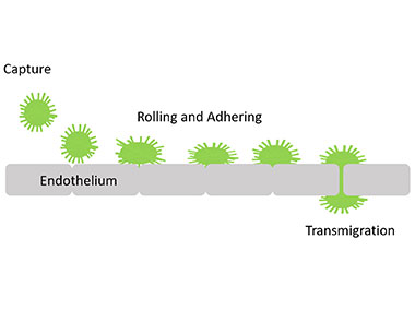
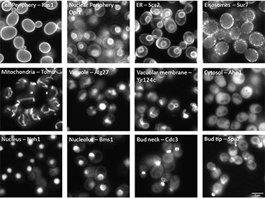
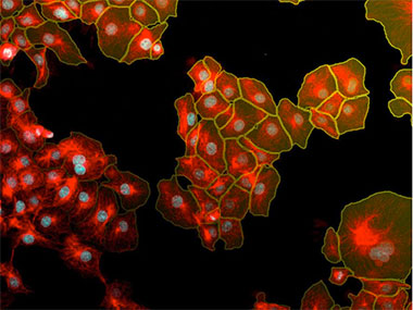
Label-Free Transmigration Assay Using scanR TruAI for Self-Learning Microscopy | Yeast Protein Localization Classified Using TruAI™ Deep-Learning Technology | Instance Segmentation of Cells and Nuclei Made Simple Using Deep Learning | 20 Examples of Effortless Nucleus and Cell Segmentation Using Pretrained Deep-Learning Models |
Related Products
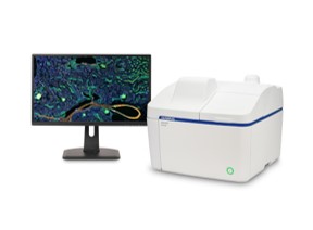
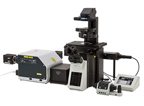
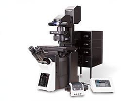
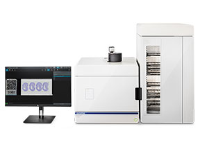
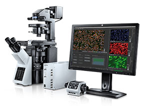
APX100
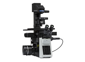
| IXplore Pro
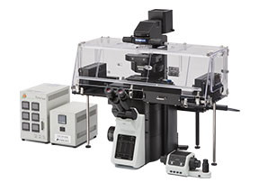
| IXplore Live
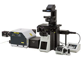
| IXplore Spin
|
IXplore SpinSR
| FV4000
*As of October 2023. | FV4000MPE
| VS200
|
scanR
|
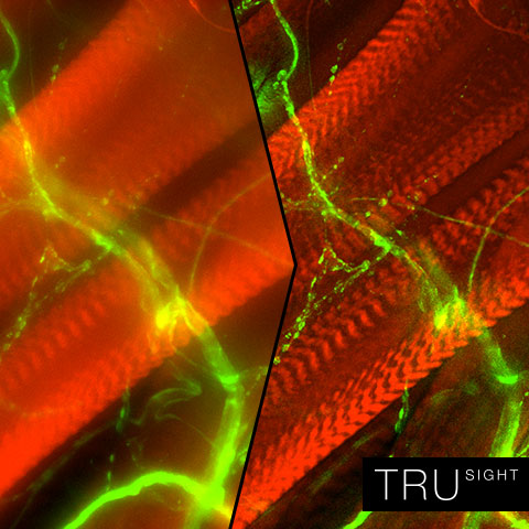
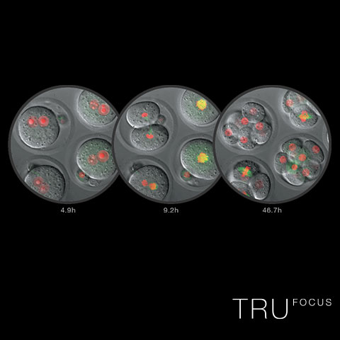
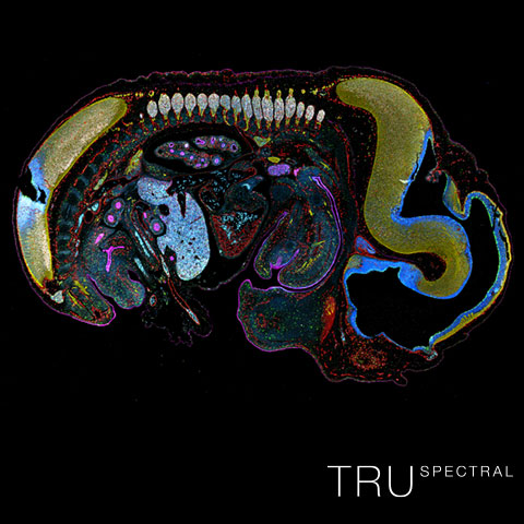
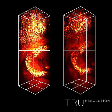
Innovative Tru technologies help researchers overcome the challenges of imaging experiments.
Seamless deconvolution for clearer, sharper images. | Stable focus throughout time-lapse experiments. | High light efficiency for bright, precise multicolor images. | Automated correction for brighter and sharper images at depth. |
Sorry, this page is not
available in your country.
Sorry, this page is not
available in your country.
.jpg?rev=5336)
.jpg?rev=36EA)


