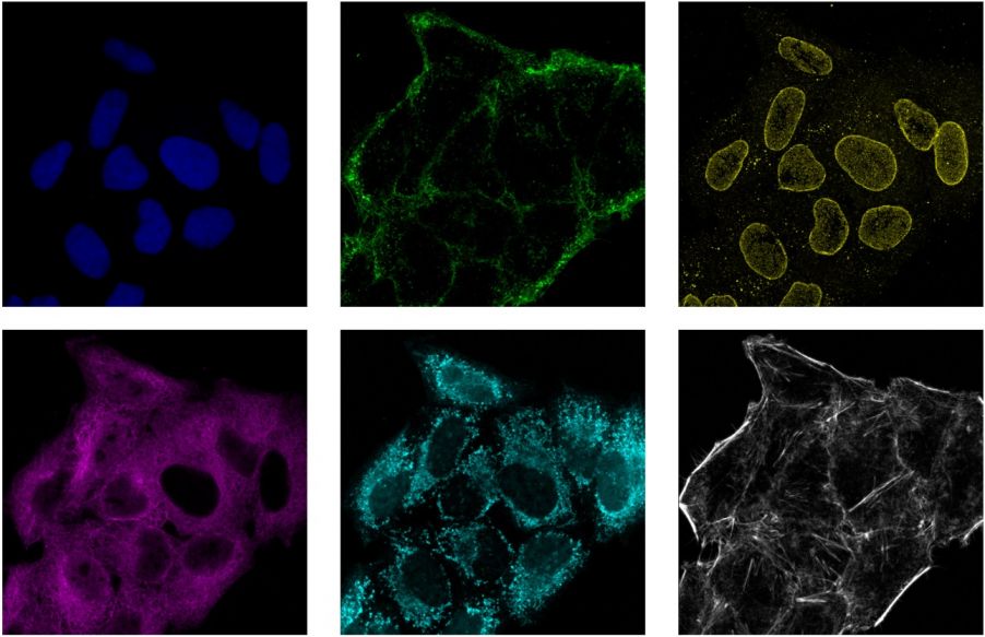
Confocal laser scanning microscopes are popular biological research tools. They're commonly used to image multiple fluorophores at once with good color separation, as well as image deep within a biological specimen with enhanced sectioning capabilities.
Today, the latest laser tech innovations can benefit applications like these and enable more advanced experiments. In this blog post, we’ll discuss how new near-infrared (NIR) excitation lasers on our FV4000 confocal microscopes are powering advanced multiplexing applications.*
Multiplexing with 5+ Channels in Confocal Microscopy – Overcoming Technical Challenges with the FV4000 System
Let’s start by casting light on the history of multiplexing experiments.
For years, many researchers performed immunofluorescence with DAPI and two other colors, typically in the green and red spectra.
As antibodies and imaging systems advanced with more detectors and better emission wavelength filtering, four-color immunofluorescence became popular. Combining DAPI, green, red, and far-red colors in one image was the most common four-color combination.
Yet, there were two critical factors that hindered the introduction of a fifth or sixth channel for multiplexing:
1. A lack of NIR laser diodes with good beam qualities
First, NIR laser diodes with good beam qualities for confocal laser scanning microscopes were not widely available. Adequate power (but not too much power), minimal power fluctuation, and compatible beam profiles are all necessary features of laser diodes used in confocal imaging. Until recently, however, only a few NIR laser diode options were available in these wavelength ranges.
But this has all changed thanks to the latest laser diode technology. Our FV4000 confocal microscope now offers 685 nm, 730 nm, and 785 nm laser diodes for efficient excitation of secondary dyes, such as those in the table below.
| Laser | Fluorescent dye | λ_Ex (nm) | λ_Em (nm) |
|---|---|---|---|
| LD685 | Alexa Fluor 680 | 679 | 702 |
| DyLight 680 | 692 | 712 | |
| Alexa Fluor 700 | 696 | 719 | |
| iRFP720 | 702 | 720 | |
| LD730 | ATTO 740 | 743 | 763 |
| DiR | 750 | 782 | |
| Alexa Fluor 750 | 752 | 779 | |
| Cy7 | 753 | 775 | |
| DyLight 755 | 754 | 776 | |
| LD785 | DyLight 800 | 777 | 794 |
| IR Dye 800CW | 778 | 794 | |
| Alexa Fluor 790 | 782 | 805 | |
| Cy7.5 | 790 | 810 |
These dyes, along with a growing number of new fluorophores, are making the addition of a fifth and sixth simultaneous channel for multiplexing more attractive.
2. Photomultiplier tubes with reduced sensitivity in NIR wavelengths
A second challenge to achieving efficient NIR imaging was that many photomultiplier tubes (PMTs), which have been used as the standard detector for laser scanning confocal microscopes, have reduced sensitivity in the detection wavelengths typical for the NIR wavelength region (above 750 nm).
This reduced sensitivity in the near-infrared detection ranges is especially true for popular GaAsP PMTs that offer higher sensitivity in the middle of the visible spectrum. In the 750+ nm range, GaAsP detectors have very low sensitivity.
To overcome this challenge, we implemented SilVIR detection technology in our FV4000 confocal laser microscope. Our SilVIR detector utilizes a silicon photomultiplier (SiPM), which is a semiconductor sensor that enables high signal-to-noise ratio fluorescence image acquisition spectrally over a wide wavelength range―from 400 nm to 900 nm.
Thanks to this technology, our FV4000 system offers up to a 6-channel SilVIR detector for multiplexing applications. Combined with our X Line™ high-performance objectives, the FLUOVEW™ FV4000 system can provide high-quality broad chromatic aberration correction from 400–1000 nm. The combination of SilVIR detectors and X-Line ™ objectives results in much better reliability in multiplexed fluorescence imaging and overall NIR imaging.
*This article was updated based on original content published on January 21, 2020.
Related Content
Brochure: FV4000 Confocal Laser Scanning Microscope
Whitepaper: Next-Generation SilVIR Detector System for the FLUOVIEW FV4000 Laser Confocal Microscope


.jpg?rev=6FBB)