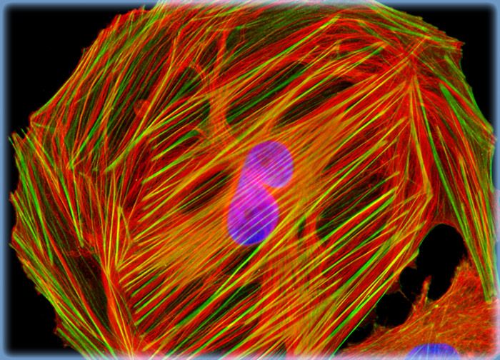
Normal African Green Monkey Kidney Fibroblast Cells (CV-1 Line)
In this section, the digital fluorescence microscopy image features CV-1 cells immunofluorescently labeled with primary anti-tubulin mouse monoclonal antibodies followed by goat anti-mouse Fab fragments conjugated to Rhodamine Red-X. In addition, the cells were stained with DAPI, which selectively binds to DNA in the cell nucleus, and Alexa Fluor 488 conjugated to phalloidin, which targets the filamentous actin network.
Sorry, this page is not
available in your country.