Judges 2010

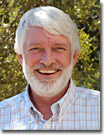
 | Dr. Douglas B. Murphy - Douglas Murphy is the Director of the Facilities for Light Microscopy, Histology and Cell Culture at the Howard Hughes Medical Institute Janelia Farm Research Campus in Ashburn, VA, and adjunct professor of cell biology at Johns Hopkins Medical School in Baltimore, MD. Dr. Murphy's past research interests in cell biology include the role of microtubules in organelle transport, the polymer dynamics of microtubules, the function of microtubule-associated proteins, and the mechanical and conformational states of the motor protein kinesin. The core facilities at Janelia Farm provide equipment and services in fluorescence and confocal imaging for laboratories throughout the research campus. The microscope facility specializes in confocal imaging, images of large tissue sections, high-throughput imaging of serial mouse brain sections, live cell imaging, and methods for measuring the dynamics of molecules in cells, including fluorescence resonance energy transfer (FRET), fluorescence recovery after photobleaching (FRAP) and total internal reflection fluorescence microscopy (TIRFM). Dr. Murphy has been a significant contributor to review articles and Java tutorials on the Olympus optical microscopy educational websites. |
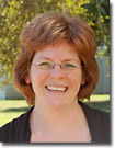
 | Dr. Catherine Galbraith - Dr. Galbraith is a senior researcher at the National Institute of Child Health and Human Development (NICHD), part of the National Institutes of Health (NIH) in Bethesda, MD, the primary agency of the United States government responsible for biomedical and health-related research. She is also currently a visiting scientist at the Janelia Farm Research Campus of the Howard Hughes Medical Institute, Ashburn, VA. Her work focuses on aspects of nanoimaging and nanotechnology, cell biology, and cellular and molecular neurobiology, including cytoskeletal dynamics and the dynamic molecular organization of structural and signaling scaffolds. A frequent instructor and leading innovator in developing and using emerging imaging technologies and new techniques for light microscopy, Dr. Galbraith employs fluorescence, polarization, confocal and superresolution microscopy techniques in her research. |
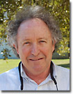
 | Dr. Mark H. Ellisman - Dr. Ellisman is Director of the National Center for Microscopy and Imaging Research (NCMIR) at the University of California, San Diego. NCMIR is a national research resource for computer-aided imaging, high voltage and intermediate voltage electron microscopy, serial section reconstruction, and functional correlation with subcellular structures in 3D and 4D. Dr. Ellisman is recognized nationally and internationally for helping to pioneer the development of new technologies that enhance neurobiological and clinical research. His own research projects include many aspects of cellular, molecular, and developmental neurobiology: mechanisms of intracellular transport in neurons; interactions between axons and myelinating glia; aging processes in the central nervous system; cellular interactions during nervous system regeneration; molecular differentiation of excitable membranes, ion channels, neurotransmitter receptors and transmembrane ion pumps; and structural changes in axons and synapses associated with changes in electrophysiological properties. |
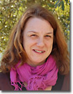
 | Dr. Anne K. Kenworthy - Dr. Kenworthy is an Associate Professor of Molecular Physiology & Biophysics, and Associate Professor of Cell and Developmental Biology, at Vanderbilt University, Nashville, TN. Her primary research interests lie in cell membrane structure, along with intracellular trafficking and protein dynamics. Her lab is using a combination of cell biology and quantitative fluorescence microscopy to explore the lipid raft hypothesis, particularly the spatial distribution and dynamics of raft-associated proteins and lipids in cells. To address these questions, she uses a variety of fluorescence microscopy techniques including fluorescence recovery after photobleaching (FRAP) and Förster resonance energy transfer (FRET). Together with biomathematicians, she is developing new methods and models with which to calibrate, measure and quantify protein dynamics in living cells. In addition, she teaches in several quantitative fluorescence microscopy courses. |
Not Available in Your Country
Sorry, this page is not
available in your country.