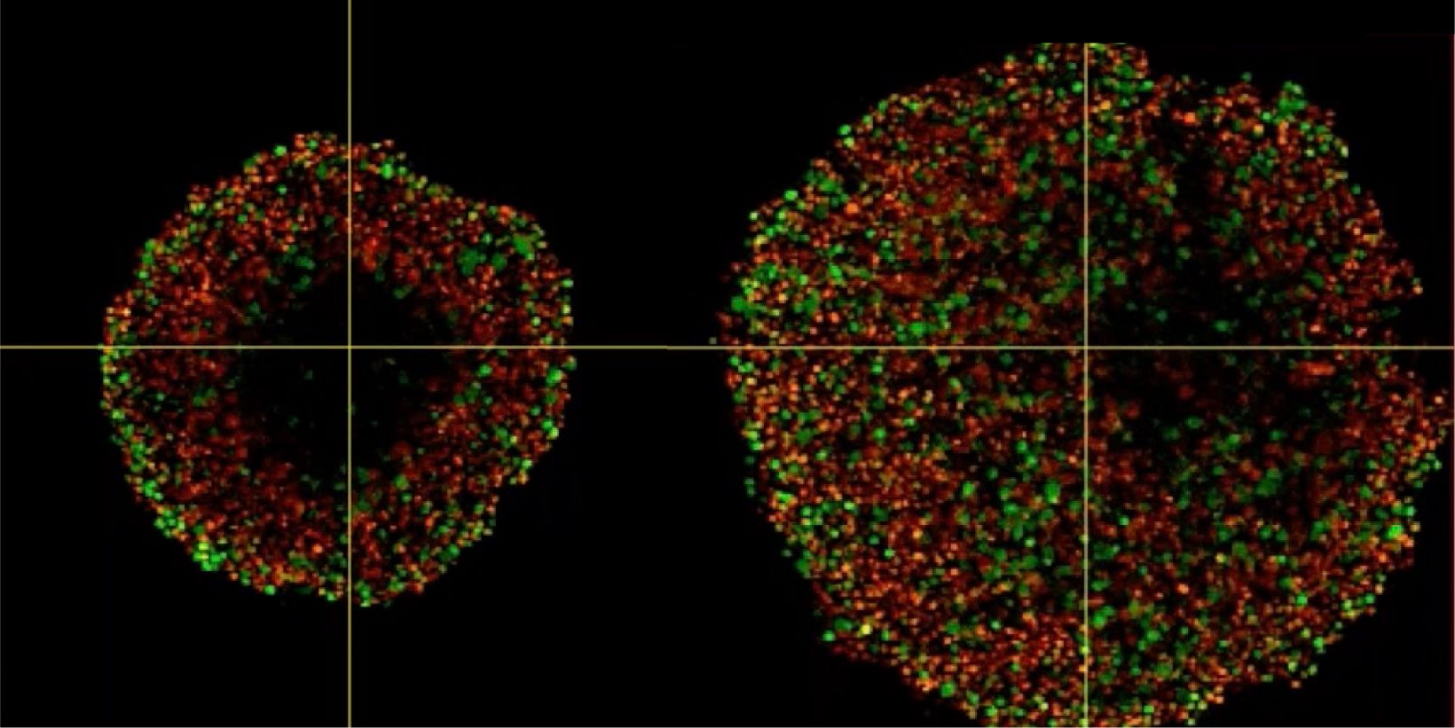
Phenotypic tumour heterogeneity arising due to differentially cycling cell populations has been implicated in increased therapy resistance. This phenomenon cannot be assessed in adherent cell culture, where microenvironmental conditions are homogeneous. Thus, we utilise melanoma spheroids to model the 3D tumour microenvironment including the extracellular matrix (ECM) and study spheroid structure, necrotic region, individual cell arrangement within and gene expression patterns. We achieve this by exploiting the fluorescence ubiquitination cell cycle indicator (FUCCI) system to monitor cell cycle stages as a surrogate marker for phenotypic tumour heterogeneity, tissue clearing and confocal microscopy using FV3000.
Investigating Spheroid Architecture Using the FV3000 Confocal MicroscopePhenotypic tumour heterogeneity arising due to differentially cycling cell populations has been implicated in increased therapy resistance. This phenomenon cannot be assessed in adherent cell culture, where microenvironmental conditions are homogeneous. Thus, we utilise melanoma spheroids to model the 3D tumour microenvironment including the extracellular matrix (ECM) and study spheroid structure, necrotic region, individual cell arrangement within and gene expression patterns. We achieve this by exploiting the fluorescence ubiquitination cell cycle indicator (FUCCI) system to monitor cell cycle stages as a surrogate marker for phenotypic tumour heterogeneity, tissue clearing and confocal microscopy using FV3000. | |
관련 제품공초점 레이저 스캐닝 현미경 FV3000
| |
Investigating Spheroid Architecture Using the FV3000 Confocal Microscope
관련 영상
관련 제품
FV3000
- 갈바노스캐너 전용(FV3000) 또는 갈바노/공진 하이브리드 스캐너 구성(FV3000RS) 에 사용 가능
- 모든 채널에 새로 개발된 정확하고 높은 효율의 TruSpectral 기술 적용
- 고감도 신호검출을 통해 낮은 광독성으로 라이브 셀 이미징 최적화

