Evident LogoOlympus Logo

Ask the Experts

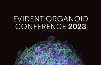
EVIDENT Organoid Conference 2023 - Looking Deeper, Capturing Complexities

Evident Organoid Conference 2023

Session 1 JeWells: A New Dedicated Organoids Platform Made for HCS and More Extreme Conditions

Session 2 3D Spheroid-Microvasculature-on-a-Chip for Tumor-Endothelium Mechanobiology Interplay

Session 3 A Sugar Code for the Dynamics of Breast Cancer Migration

Session 4 Complex In Vitro Tumor Models and 3D Confocal Imaging Technology to Investigate Cell Therapy against Solid Tumor


Agenda

Time (GMT +8) February 8, 2023

11:00 a.m. - 11:10 a.m.

Opening Address by Akio Hirohashi
Member of Board, Chief Strategy Officer, Evident Corporation

Chairperson
Dr. Graham Wright

Director, Research Support Centre (RSC)
A*STAR, Singapore

11:10 a.m. - 11:55 a.m.

Session 1
JeWells: A New Dedicated Organoids Platform Made for HCS and More Extreme Conditions

Presenter:
Dr. Anne Beghin

Research Assistant Professor
National University of Singapore

More information

11:55 a.m. - 12:40 p.m.

Session 2
3D Spheroid-Microvasculature-on-a-Chip for Tumor-Endothelium Mechanobiology Interplay

Presenter:
Dr. Arnold Ju

School of Biomedical Engineering,
Faculty of Engineering and Sydney Nano Institute
The University of Sydney, Australia

More information

12:40 p.m. – 1:25 p.m.

Session 3
A Sugar Code for the Dynamics of Breast Cancer Migration

Presenter:
Dr. Ramray Bhat

Assistant Professor
Indian Institute of Science, Bangalore, India

More information

1:25 p.m. – 2:10 p.m.

Session 4
Complex In Vitro Tumor Models and 3D Confocal Imaging Technology to Investigate Cell Therapy against Solid Tumor

Presenter:
Dr. Fuqian Xie

Cofounder and Chief Scientific Officer
Shanghai Biocast Biotechnology Co. Ltd., Shanghai, China

More information

2:10 p.m. – 2:55 p.m.

Session 5
Genotype-Phenotype Association Strategy for Fatty Liver Diseases Using Human iPSC-Derived Organoids

Presenter:
Dr. Yosuke Yoneyama

Assistant Professor
Tokyo Medical and Dental University, Japan

More information

2:55 p.m. – 3:00 p.m.

Closing Address by Jian Shen
General Manager, Life Science
Evident APAC


Speakers

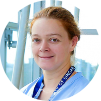
Dr. Anne Beghin

Assistant Professor, Research
Mechanobiology Institute, National University of Singapore

Dr. Arnold Ju

School of Biomedical Engineering,
Faculty of Engineering and Sydney Nano Institute
The University of Sydney, Australia

Dr. Anne Beghin

Biography

Dr. Anne Beghin is a multidisciplinary scientist with 15 years of extensive research experience across academia and industry. She obtained her PhD in oncology in 2007 at the University Claude Bernard in Lyon, France. She then moved to optical microscopy at the Université de Lyon, where she established the microscopy platform and developed live-cell imaging solutions and image analysis services for four years.

In 2011, she was recruited by a biotechnology company based in Bordeaux, where she spent three years in charge of a tissue analysis service: from biologic samples (whole tissue sections and tissue microarrays) to image acquisition and analysis with database establishment. She has been part of the Interdisciplinary Institute of Neuroscience (IINS) for three years, where she successfully developed a new platform linking the high-content screening (HCS) approach with super-resolution microscopy such as single-molecule light microscopy (HCS-SMLM), a collaboration with the pharmaceutical company, Sanofi. She then moved to the Mechanobiology Institute (MBI) in Singapore to study organoids using advanced imaging and HCS. This work has resulted in a patent and ongoing publications.

Abstract

JeWells: A New Dedicated Organoids Platform Made for HCS and More Extreme Conditions

Turning organoids into an impactful translational technique includes being able to generate and assess organoids that develop robustly with physiologically relevant architecture. However, quantitative comparisons and statistical analysis at high content, which are mandatory to describe the complexity of such multicellular 3D objects, are hindered by the lack of high-throughput 3D imaging methods.

As a result, we engineered a versatile high-content screening (HCS) device to streamline the steps of organoids and organotypic cultures to exploit their potential in morphogenesis and tissue homeostasis understanding. Our approach comprises a new generation of versatile scaffolding cell culture multiwell chips with embedded optical components (= lighting JeWells) that enables fast 3D imaging. Biofunctionalization of JeWells with extracellular matrix (ECM) components produces precise microniches to grow and differentiate microphysiological elements.

The device is fully compatible with classical imaging techniques such as brightfield, widefield, light sheet, or confocal microscopy. The defined positioning of organoids enables correlations between all these different techniques without any loss of organoids. The high surface density of JeWells meets HCS standards: we can generate more than one hundred organoids on the surface equivalent to a single well of a 384-well plate.

Our platform enables one to follow the morphogenesis of living organoids over weeks using non-toxic fluorescent live imaging based on light sheet microscopy (validated on human embryonic stem cells (hESC), human-induced pluripotent stem cells (hIPSC), and primary cells). The possibility to build versatile and tunable 3D microniches with the ECM inside the JeWells opens the ability to use these chips on organotypic cultures and microphysiological systems.

The large number of 3D images can be used to train convolutional neural networks to precisely detect and quantify subcellular and multicellular features, such as mitotic and apoptotic events, multicellular structures (rosettes), and classify whole organoid morphologies. Combining high-resolution 3D microscopy techniques with HCS and machine learning approaches enables us to quantitatively describe the morphogenesis of hundreds of living organoids correlated with the phenotypic characterization to decipher mechanisms involved in morphogenesis and human physiopathology, as well as the response to extreme conditions.

Dr. Arnold Ju

Biography

Arnold Ju received his PhD in biomedical engineering at the Georgia Institute of Technology and Emory University in Atlanta, Georgia, USA. In 2014, he joined the Australian Centre for Blood Diseases at the Monash University in Melbourne, Australia, as a junior postdoc. He relocated in 2015 to Sydney, Australia, to join the Heart Research Institute. In early 2020, Dr. Ju joined the University of Sydney (USYD)’s new biomedical engineering school as a senior lecturer and started up the Mechanobiology and Biomechanics Laboratory (MBL).

Dr. Ju currently holds a Heart Foundation Future Leader Fellowship, working at the interface between mechanical engineering and mechanobiology. His team has pioneered multiple biomechanical nanotools, including blood clot-on-chip microfluidic devices (Nature Materials 2019), single-cell biomembrane force probes (Nature Communications 2018), and 4D haemodynamic modeling (Nature 2021).

His vision is to build novel platforms that integrate advanced biomanufacturing, high-throughput biomechanical manipulation, and artificial intelligence for biobank data processing. His track record spans developing, characterising, and evaluating innovations of 3D organoids and organ-on-chips, mechanobiology, imaging probes and biosensors, bio-nanotechnology, and image-based deep learning. These large facilities should provide significant benefits to interdisciplinary research in biofabrication, biomechanics, and point-of-care microtechnologies.

Abstract

3D Spheroid-Microvasculature-on-a-Chip for Tumor-Endothelium Mechanobiology Interplay

In the final step of cancer metastasis, tumor cells become lodged in a distant capillary bed, where they can undergo extravasation and form a secondary tumor. While increasing evidence suggests blood/lymphatic flow and shear stress play a critical role in the tumor extravasation process, there is a lack of systematic and biomechanical approaches to recapitulate sophisticated 3D microtissue interactions within the controllable hydrodynamic microenvironment.

Here, we report a simple-to-use 3D spheroid-microvasculature-on-a-chip (SMAC) model. Under static and controlled flow conditions, the SMAC recapitulates the biomechanical crosstalk between heterogeneous tumor spheroids and the endothelium in a high-throughput and quantitative manner. As an in vitro metastasis mechanobiology model, we discover 3D spheroid-induced endothelial compression and cell-cell junction degradation in the process of tumor migration and expansion. Lastly, we examine the shear stress effects on the endothelial orientation, polarization, as well as the tumor spheroid expansion.

Taken together, our SMAC model offers a miniaturized, cost-efficient, and versatile platform for future investigation on metastasis mechanobiology, enhanced permeability and retention effect, and even personalized therapeutic evaluation.

Dr. Ramray Bhat

Assistant Professor
Indian Institute of Science, Bangalore, India

Dr. Fuqian Xie

Cofounder and Chief Scientific Officer
Shanghai Biocast Biotechnology Co. Ltd., Shanghai, China

Dr. Ramray Bhat

Biography

Ramray Bhat is an associate professor in the department of Molecular Reproduction Development and Genetics, and an associate faculty with Biosystems Science and Engineering at the Indian Institute of Science in Bangalore, India. He is a member of the EMBO Global Investigator Network. He holds an undergraduate degree in medicine from the University of Calcutta and a PhD in cell biology and anatomy from the New York Medical College. He was a Komen postdoctoral fellow at the Lawrence Berkeley National Laboratory. His interests are in cancer, development, and evolution, and his group’s research is funded by the Wellcome Trust-DBT India Alliance and the John Templeton Foundation, USA.

Abstract

A Sugar Code for the Dynamics of Breast Cancer Migration

Carcinogenesis is a process where frameworks of interactions between untransformed cells and their surroundings are subverted and replaced by new frameworks that seek to drive canonical behaviors of cancer cells. One such behavior is their tendency to migrate through surrounding extracellular matrices in a bid to spread to other organs of the body, a process known as metastasis. Migrations can be complex and of distinct types. They are functions of the genetic and epigenetic aberrations that contribute to the new interactive frameworks associated with cancer cells. Alterations in the levels of glycans and their binding proteins, lectins are among the oldest and ubiquitous epigenetic changes in oncology.

I will showcase how we built a multiscale computational model that predicts the structure of these frameworks, as well as the diversity of cancer migration modes. I will then discuss two short stories from my group, where alterations in levels of the galactoside-binding protein galectin-9 on the one hand, and intercellular heterogeneity in levels of 2,6-linked sialic acids on the other, drive distinct modes of migration of triple-negative breast cancer cells. Our mechanistic findings reveal glimpses of a glycocode that could underlie the biophysical phase transitions in the migratory behavior of breast cancer.

Dr. Fuqian Xie

Biography

Fuqian Xie received his PhD in biophysics in 2008, then completed post-doctoral training at the Washington University School of Medicine in St. Louis, Missouri, USA. His research fields and accomplishments include the mechanistic study of DNA repair, anti-viral drug screening with imaging-based assay development, microfluidics, total internal reflection fluorescence (TIRF), and single-molecule spectroscopy.

After joining a New York-based biotech company in 2013, he played a key role in leading and completing preclinical studies of several biological candidates using complex in vitro models in research and development (R&D) pipelines that enabled Investigational New Drug (IND) submissions targeting nonalcoholic fatty liver disease (NAFLD) and central nervous system (CNS) diseases.

In 2017, he cofounded Shanghai Biocast Co. Ltd. as the Chief Scientific Officer. He currently leads a passionate team of specialists and scientists using various confocal microscope platforms and their bio-imaging expertise to tackle problems in drug R&D, including imaging-based phenotypic assay development, complex 3D disease modeling, and spatial omics in translational or clinical stages of drug development.

Abstract

Complex In Vitro Tumor Models and 3D Confocal Imaging Technology to Investigate Cell Therapy against Solid Tumor

Cell therapy genetically engineered with chimeric antigen receptors (CARs) have been successful in treating hematological malignancies, while treatment for solid tumors have been modest. Researchers are attempting novel optimization and quantifying strategies to improve clinical outcomes in patients treated with cell therapy. Factors in solid tumor that hinder the efficacy in cell therapy include difficulty with infiltration to the tumor and the complex nature of its microenvironment, such as the extracellular matrix (ECM), stroma cells, and immune cells. A 3D in vitro tumor model is a translational tool that can improve predictions of the clinical efficacy of cell therapy.

Here we created 3D spheroid and patient-derived tumoroids models using various methods in a high-throughput format to recapitulate a 3D structure of a solid tumor and an in vivo-like tumor microenvironment (TME). We demonstrated that a 3D confocal imaging system enables the monitoring of 3D tumor models in a high-throughput manner and that it performs spatial and volumetric imaging analysis. Cocultures of 3D tumor models and chimeric antigen receptor natural killers (CAR-NKs) specifically targeting carcinoembryonic antigen (CEA) on tumor cells could be imaged over a long period. Fluorescence profiling over time indicated potency and efficacy of cell therapy, such as NK cytotoxicity, NK infiltration, and antibody-dependent cellular cytotoxicity (ADCC).

The volumetric analysis of tumoroids and spatial statistics profiling between NKs and tumors offer an abundance of qualitative and quantitative data describing the killing and infiltration events of NKs in a model of a solid tumor. These imaging-based assays empowered us to determine a more comprehensive CAR-NK profile and identify highly effective immunotherapeutic agents versus multi-parametric analysis.

Dr. Yosuke Yoneyama

Assistant Professor
Tokyo Medical and Dental University, Japan

Dr. Yosuke Yoneyama

Biography

Yosuke Yoneyama obtained his PhD from the University of Tokyo in Japan, where he continued his post-doctoral work on spatiotemporal control of the growth factor signaling system. He then joined the laboratory of Dr. Takanori Takebe at the Tokyo Medical and Dental University in Japan as an assistant professor. He now focuses on the basic biology of pluripotent stem cells at the molecular and cellular levels, and works on the metabolic diseases, including fatty liver diseases by using human organoid models.

Abstract

Genotype-Phenotype Association Strategy for Fatty Liver Diseases Using Human iPSC-Derived Organoids

Nonalcoholic fatty liver disease (NAFLD) is the most common liver disease worldwide, and a major risk for cirrhosis and hepatocellular carcinoma. Earlier prediction of pathogenic NAFLD is a public health priority yet remains challenging due to the complex disease contexts affected both by genetic pleiotropy and metabolic disorders.

Recently, we developed a human population organoid panel strategy for genotype-phenotype association studies of fatty liver diseases. To qualify and control the organoid manufacturing, we set up the combination of a live-cell monitoring system and biochemical assays, leading to the reproducible protocol for generating human liver organoids from the induced pluripotent stem cells (iPSCs) of multiple donors. Fatty liver organoids derived from human iPSCs of multiple genotyped individuals enabled us to efficiently evaluate the association of particular gene variants with NAFLD phenotypes in a dish.

We propose that metabolically resolved genetic and phenotypic assessments are critical to identify biomarkers and tailor interventional strategies at a personalized level.


관련 제품

배양 모니터링 시스템

CM20

  • 정량 데이터 자동 측정
  • PC 또는 태블릿에서 원격으로 배양 진행 상황 모니터링
  • 소형 디자인용 사선 에피 조명을 갖춤
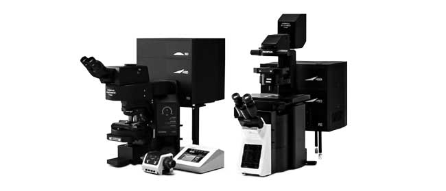
컨포칼 레이저 스캐닝 현미경

FV3000

  • 검류계 전용(FV3000) 또는 검류계/공진(FV3000RS) 하이브리드 스캐너 구성에 사용 가능
  • 모든 채널에 정확한 고효율 TruSpectral 검출
  • 고광도 및 저광독성으로 라이브 세포 이미징 최적화
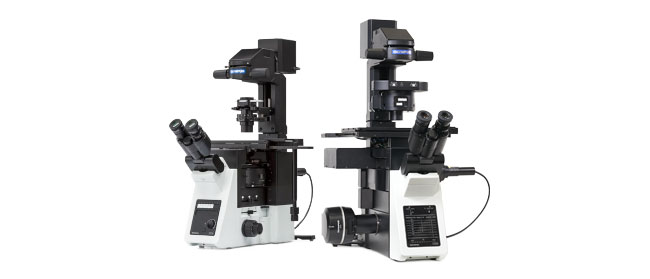
Inverted Imaging Platform

IXplore Series

  • Designed to help by providing solutions-based packages that suit your research applications
  • Accurate, reproducible images and data in a system that can be adapted as your experiment needs evolve or become more complex over time
3D 세포 분석 소프트웨어

NoviSight

NoviSight 3D 세포 분석 소프트웨어는 마이크로플레이트 기반 실험에서 회전 타원체 및 3D 물체에 대한 통계 데이터를 제공합니다. 이 소프트웨어를 사용하면 세포 활동을 3D로 정량화하고, 희귀한 세포 이벤트를 쉽게 포착하며, 정확한 세포 개수를 하고, 검출 감도를 향상할 수 있습니다. NoviSight 소프트웨어는 포인트 스캔 공초점 이미지화, 2광자 이미지화, 스피닝 디스크 공초점 이미지화, 초고해상도 생세포 이미지화 등 다양한 이미지화 방법을 적용합니다.

  • 전체 구조에서 세포 이하 기능에 이르기까지 빠른 3D 이미지 인식
  • 정확한 통계 분석
  • 바로 사용 가능한 기본 분석이 다양하게 제공되며 손쉬운 맞춤 설계 또한 가능
EVIDENT Organoid Conference 2023 - Looking Deeper, Capturing Complexities2024년11월6일
죄송합니다. 이 페이지는 해당 국가에서 사용할 수 없습니다.
Ask the Expert Sign-up

By clicking subscribe you are agreeing to our privacy policy which can be found here.

Sorry, this page is not
available in your country.

죄송합니다. 이 페이지는 해당 국가에서 사용할 수 없습니다.