Three-Dimensional High-Throughput Image Analysis of Patient-Derived Organoids and Spheroids
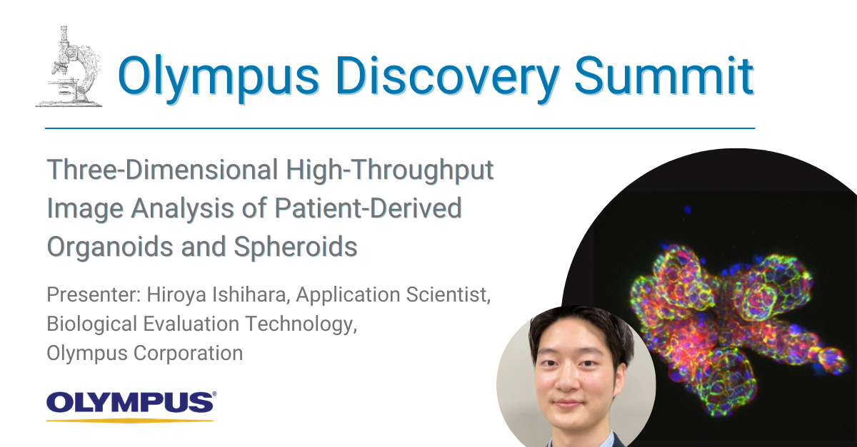
Organoids and spheroids can more faithfully reproduce in vivo conditions, and imaging-based analysis can monitor cell-specific responses with high spatial resolution. Therefore, we have been developing imaging-based three-dimensional analysis and drug evaluation methods using patient-derived cancer organoids and spheroids. In this presentation, we discuss examples of applications with recent trends in organoid research.
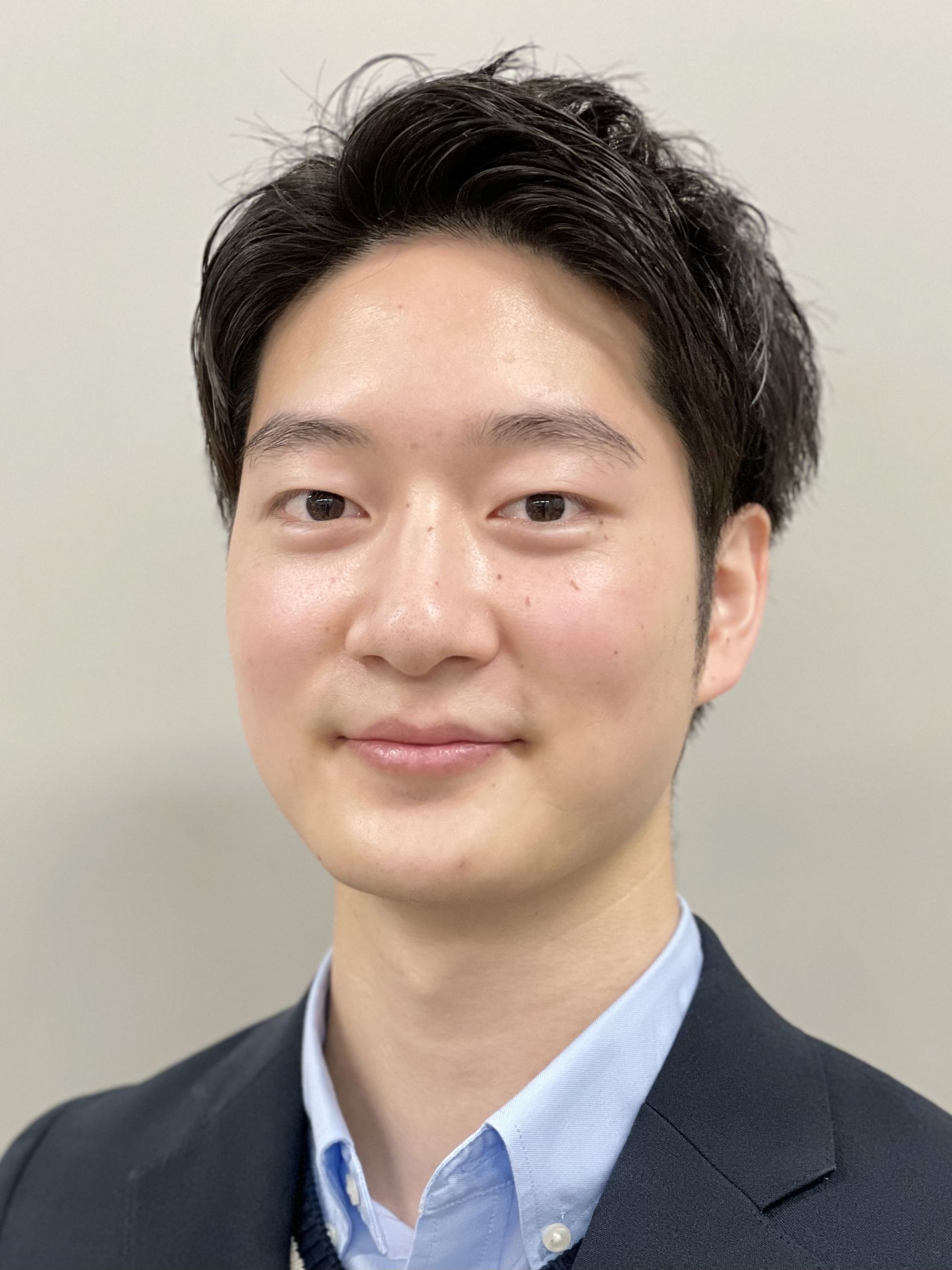
Presenter: Hiroya Ishihara, Application Scientist, Biological Evaluation Technology, Olympus Corporation
Hiroya Ishihara is an application scientist at Olympus Tokyo. His time studying the epigenetic factors involved in plant regeneration using omics and microscopy and using confocal, two-photon microscopy in that study, motivated Hiroya to join Olympus to make life science more exciting with microscopes. Currently, he is working on a wide range of projects from basic research to product and sales strategy.
Three-Dimensional High-Throughput Image Analysis of Patient-Derived Organoids and Spheroids
|
このページはお住まいの地域ではご覧いただくことはできません。

