Polarized Microscopy and What It Can Teach Us About the Materials That Make Up Our Skeletal Tissue
Introduction
Polarized microscopy is an advanced optical technique that enhances contrast in birefringent specimens. It offers distinct insights into the structure and composition of a wide range of organic and inorganic materials.
Two types of polarized microscopy are commonly used: linear polarization and circular polarization. As its name implies, linear polarized microscopy uses linearly polarized light to illuminate the specimen. In contrast, circular polarized microscopy employs circularly polarized light, making it particularly sensitive to the chirality (left or right orientation) of structures within the specimen. These techniques are used in fields such as biology, materials science, and geology for examining the microscopic structure and properties of various materials.
This application note explores these polarized light techniques in detail, highlighting how circular polarized microscopy enables better visualization of the birefringent materials in skeletal tissue.
The Birefringent Materials of Skeletal Tissue
Birefringence is the property of a material where a single incident light ray splits into two distinct rays when it passes through the material. The skin, cornea, tendons, ligaments, muscle tissue, cartilage, and bone are all tissues and organs that exhibit birefringence because of their organized, anisotropic structures (optical properties are not uniform in all directions). This birefringence affects how the materials in our skeletal tissue, such as bones and cartilage, interact with polarized light.
Bones in mammals and birds are stiff materials principally made of collagen and calcium phosphate. The four cell types found in bones are:
- Osteoblasts, which build new bone.
- Osteocytes, which are embedded in the bone matrix.
- Osteoclasts, which reabsorb bone.
- Osteoprogenitor cells, which give rise to osteoblasts.
Cartilage works together with the bone to make what it is known as skeletal tissue. This specialized connective tissue is flexible but tough. The three different types of cartilage are:
- Hyaline cartilage: the most common type found throughout the body, including the nose, ribs, trachea, larynx, bronchi, and articulating surfaces of bones. It plays an important role during embryonic development and growth by forming the fetal skeleton and growth plate, which is later replaced by bone.
- Fibrocartilage: found between the vertebrae of the spine and in some joints.
- Elastic cartilage: found in the epiglottis, vocal cords, and external ear.
Many substances are known to have an effect in bone and cartilage health. One is vitamin C, also known as ascorbic acid. Scientific studies have shown that vitamin C is essential for bone health because it is required for collagen formation and it can induce the expression of bone matrix genes in osteoblasts. In cartilage, vitamin C has been associated with slowing osteoarthritis (a disease that causes articular degeneration) because it stimulates collagen synthesis.
Understanding Linear and Circular Polarized Microscopy
To capture high-quality images of bones, cartilage, vitamin C, and other birefringent materials using polarized microscopy, it is important first to understand the linear and circular polarized lighting methods.
Light sources generate unpolarized light, which vibrates in all 360-degree angles. When unpolarized light passes through a polarizer, it is converted into linear polarized light. The linearly polarized light passes through the sample. If the sample is isotropic (uniform optical properties in all directions), it will not affect the polarized light, and the light will remain in the same polarization state. However, if the sample is anisotropic, it will change the polarization of the light as it passes. This change in polarization enables the light to pass through an analyzer, which is oriented perpendicular to the polarizer.
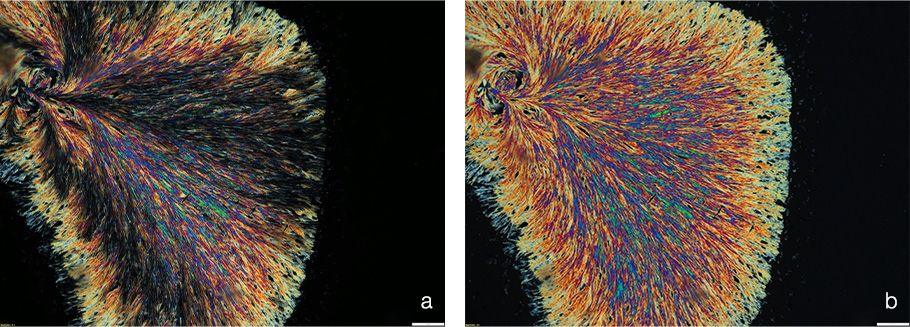
A drawback of linear polarization is the formation of isogyres, dark bands in a Maltese cross pattern (cross with V-shaped arms) that appear in the field of view. This occurs when the sample imaged has radial symmetry, which causes the polarized light to split around the radial center and create light that cannot pass through the analyzer. The Maltese cross causes a reduction of intensity, so it affects the use of the images for quantification or analysis.
Circular polarization does not have this drawback. It also uses a polarizer to convert ordinary light into light vibrating in a single plane (linearly polarized light). However, in circular polarization, a birefringent material such as a quarter-wave plate is placed at a 45° angle relative to the polarizer in the path of the polarized light. Light will experience a phase shift, which is the difference in the time it takes for the light waves to pass through the material. This results in light with a circular rotating field. When the circular rotating light passes through the specimen, it is refracted in all 360-degree rotational positions. By placing a second quarter-wave plate at a 90° angle from the first plate in the path of the refracted light, this effect will cancel. This creates linear polarized light again, which is then allowed to pass through the transmission axis of the analyzer. It is important to state that both quarter-wave plates are rotated by 45° angles with respect to the polarizer but in opposite directions.

Figure 1. Vitamin C crystals imaged with a) linear and b) circular polarized microscopy using the SLIDEVIEW™ VS200 research slide scanner by Evident with the MPLFLN40X (0.75 NA) objective. (a) With linear polarization, we can see the characteristic isogyres (black bands).
(b) With circular polarization on the same sample, we cannot see any artifacts.
Imaging Bone, Cartilage, and Vitamin C Using Polarized Microscopy
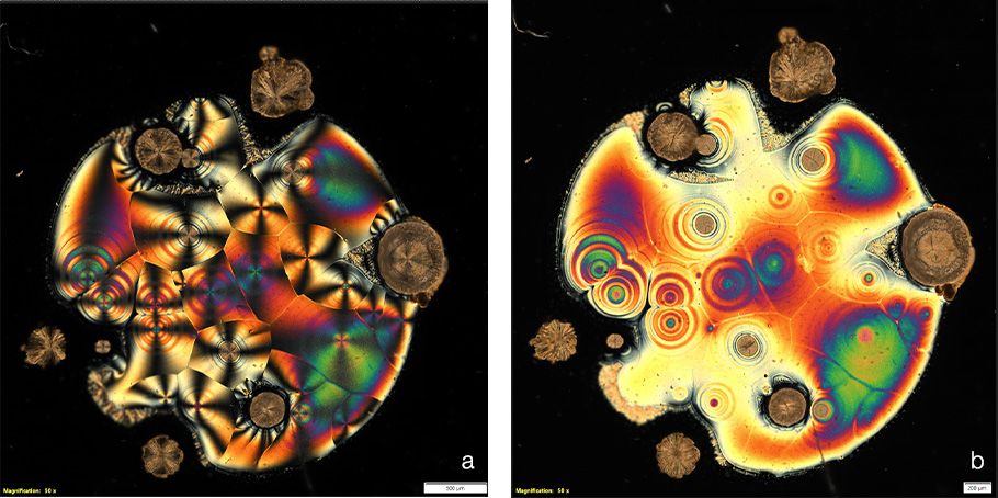
Vitamin C, also known as ascorbic acid, is a chiral molecule. When polarized light passes through vitamin C crystals, the molecule's chirality causes the rotation of the light’s polarization plane. The light interference produces a range of colors, leading to beautiful images.

Figure 2. Images of vitamin C under polarized microcopy. a) With linear polarization, we can see the characteristic isogyres (Maltese crosses). b) Circular polarization does not produce the Maltese crosses. The brown stone-like structures shown here are very thick crystals. Images captured using the VS200 research slide scanner with the MPLAPON50X (0.95 NA) objective.

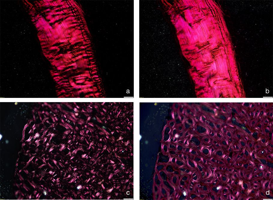
Figure 3. Images of connective tissue and bone, where collagen fibers of differing orientation are visualized in polarized light. a) Connective tissue visualized using linear polarized light. b) Connective tissue visualized using circular polarized light. c) Transverse bone section visualized using linear polarized light. d) Transverse bone section visualized using circular polarized light. Collagen fibers aligned transversely appear bright, while those aligned longitudinally appear dark. Fibers with intermediate orientations show varying shades of gray. Images captured using the VS200 research slide scanner with the MPLAPON50X (0.95 NA) objective.
The Importance of Using Polarized Imaging for Skeletal Tissue and Vitamin C Studies
Capturing beautiful images using polarized microscopy is aesthetically pleasing, but what is the scientific relevance of the images? In the case of vitamin C, the interaction of the molecules with polarized light is a property used in chemical analysis to determine the concentration and purity of vitamin C. This is normally done using a polarimeter.
In bone, studies using circular polarized microscopy have been used to map collagen fiber orientation patterns. This has been correlated to bone strain data. It is now known that collagen fibers in a predominantly transverse orientation have better resistance to compressive forces, while longitudinal fibers have better resistance to tensile forces. In addition, collagen fibers oriented at 45° angles from the lamella provide better resistance to shear.
The orientation of collagen fibers in cartilage has also been shown to be important for the resistance of pressure and deformation due to loading or motion. Disruptions in the collagen organization within cartilage, even if minor, have been linked to pathological diseases such as osteoarthritis.
*Circular polarization components for the VS200 scanner are currently only available in the EMEA. Please contact your local Evident sales representative for details on their availability.
References
- Bromage, T., et al. 2023. "Circularly Polarized Light Standards for Investigations of Collagen Fiber Orientation in Bone." The Anatomical Record. 274(1): 157–168.
- Chin, K. Y., and I-N. Soelaiman. 2018. "Vitamin C and Bone Health: Evidence from Cell, Animal, and Human Studies." Current Drug Targets. 19(5): 439–450.
- Aghajanian. P., et al. 2025. "The Roles and Mechanisms of Actions of Vitamin C in Bone: New Developments." Journal of Bone and Mineral Research. 30(11): 1945–1955.
- Khebtsov, N., et al. 2016. "Chapter 1: Introduction to Light Scattering by Biological Objects." Handbook of Optical Biomedical Diagnostics. 2nd ed., vol. 1: Light-Tissue Interaction, edited by V. V. Tuchin.
- Xia. Y., et al. 2016. "Chapter 1: Introduction to Cartilage." Biophysics and Biochemistry of Cartilage by NMR and MRI. Royal Society of Chemistry. 1–43.
- Clark. A., et al. 2002. "The Effects of Ascorbic Acid on Cartilage Metabolism in Guinea Pig Articular Cartilage Explants." Matrix Biology. 21(2): 175–184.
- Mittelstaedt. D., et al. 2011. "Quantitative Determination of Morphological and Territorial Structures of Articular Cartilage from Both Perpendicular and Parallel Sections by Polarized Light Microscopy." Connective Tissue Research. 52(6): 512–522.
Authors
Laura Lleras Forero, Product Marketing Manager, Life Science Research, EMEA, Evident
Heiko Gäthje, Senior Trainer, Training Academy, Evident
このアプリケーションノートに関連する製品
Maximum Compare Limit of 5 Items
Please adjust your selection to be no more than 5 items to compare at once
このページはお住まいの地域ではご覧いただくことはできません。