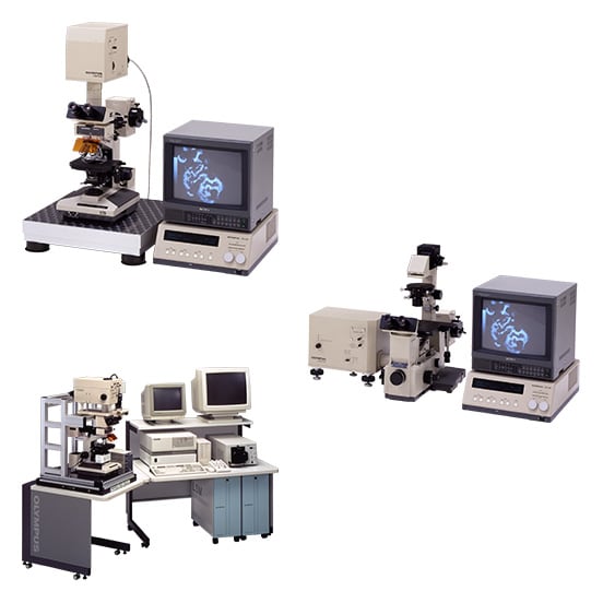
Scanning microscopes have long been researched because their superior resolution and contrast allows for the construction of optical cross-sectional images (3D images). Moreover, the models are unique in that a light stimulus can be applied to the specimen. In the 1970s, Olympus announced a semiconductor-testing device that used photoexcited current observations. The company also continued developing laser technologies. After a period when this work was suspended, Olympus, in 1983, engineered a confocal microscope capable of constructing 3D images. The company then ran repeated tests on its prototype laser scanning microscope. In 1990, Olympus developed the upright LSM-GB and the inverted LSM-GI for biology applications. The upgraded LSM-GB200 was launched in 1992. All these microscopes used the BHS microscope body. The LSM-GB featured an image memory capacity that could hold two 640 × 480-pixel 8-bit images. Confocal laser scanning microscopes were supported by cutting-edge technologies, including rapid optical scanning, weak light detection and photoelectric conversion (photon counting), as well as an optical glass filter with accurate wavelength selectivity (precise, multilayer film coating technology). Today, laser scanning microscopes are becoming faster and producing higher resolution images as computers become more advanced. |
Not Available in Your Country
Sorry, this page is not
available in your country.
You are being redirected to our local site.