Trust the Colors with Olympus True Color LED
True Color LED illumination is a durable, bright light source with spectral properties that closely match halogen lamp illumination. Olympus’ market-leading LED technology delivers accurate color reproduction that provides the confidence needed for reliable diagnoses in pathology.
Halogen lamp illumination has for many years been the gold standard in microscope illumination. The main reason is that thanks to its particular properties, excellent color reproduction – both through the oculars and on the computer screen – can be obtained.
Today, LED technology has become a viable alternative for conventional lighting in many areas of everyday life. Its bright, efficient light output provides many benefits across different industries. However, in the field of microscopy, where incorrect color reproduction can impact the evaluation of a sample, LED technology shows its shortcomings, often introducing undesired color shifts. So, what could make an LED suitable for the rigorous demands of clinical microscopy?
Measuring the Quality of a Light Source
An LED light source for microscopy should emit white light, but should also be able to match or even surpass the capability of halogen lamp light to show the colors of the sample correctly. How has the halogen lamp light source up until now been able to perform well at this task? It turns out that the key is its mostly even intensity across the visible spectrum, which allows uniform illumination of all the different colors in a sample (figure 1).
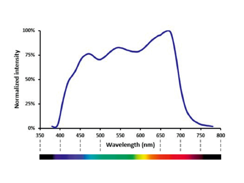
Figure 1: White Light
The spectrum of a halogen lamp light source shows its even intensity across the visible spectrum when correctly set at 9 V with a daylight filter.
At a Glance
- Generic LED light sources provide low energy consumption and a long lifetime, but usually alter the colors of a stained sample.
- Olympus’ True Color LED solves this issue by employing dedicated technologies that avoid color distortion and provide reliable images.
Trust the Colors with Olympus True Color LED
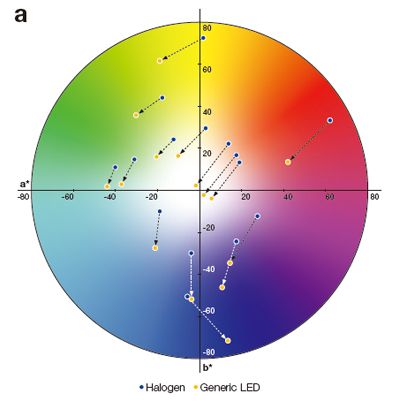
Different light sources each have a unique illumination spectrum. While it is obvious that differences in spectra will alter how sample colors are represented, a precise and quantifiable comparison of light sources reveals the true impact of such variation. One way of making this comparison in a standardized, reproducible manner, is to represent the rendered colors on a chromaticity diagram. With a chromaticity diagram, such as the a*b* chart in figure 2, it becomes easy to visually compare the color rendering capabilities of two different light sources, showing the impact of the light source’s spectrum. Spectral differences between light sources will result in the same reference colors appearing at different locations in the diagram.
One way of making this comparison in a standardized, reproducible manner, is to represent the rendered colors on a chromaticity diagram. With a chromaticity diagram, such as the a*b* chart in figure 2, it becomes easy to visually compare the color rendering capabilities of two different light sources, showing the impact of the light source’s spectrum. Spectral differences between light sources will result in the same reference colors appearing at different locations in the diagram.
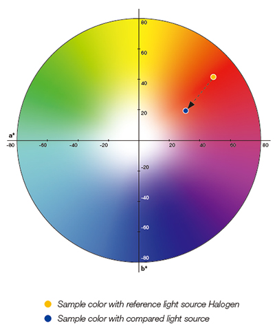
Figure 2: Chromaticity Diagram
Color shifts can be shown in an intuitive, visual way in a*b* charts. In this case the compared light source heavily shifts the sample color towards a more blueish hue.
Matching Colors
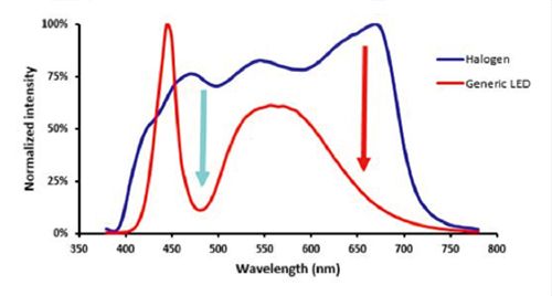
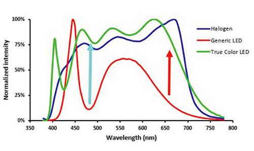
Many LED designs have so far not produced the light quality that is required for reliably distinguishing colors in a specimen. The reason behind this becomes clear when comparing the emission spectra of the two light sources (figure 3).
Compared to the halogen lamp spectrum generic LEDs show more variability, especially at 480 nm and in the region between 600 nm and 700 nm. These wavelengths correspond to histologically important colors. Having an uneven relative intensity is a generic problem induced by LED illumination – and this variability has a clear effect on the color rendering ability of an LED light source.
Figure 3: Halogen lamp vs LED
The intensity variation of the LED’s spectrum leads to low relative illumination in the cyan and red regions (arrows).
Compared to the halogen lamp spectrum generic LEDs show more variability, especially at 480 nm and in the region between 600 nm and 700 nm. These wavelengths correspond to histologically important colors. Having an uneven relative intensity is a generic problem induced by LED illumination – and this variability has a clear effect on the color rendering ability of an LED light source.
The effect of such a significantly different illumination spectrum can be clearly seen in a chromaticity diagram. Figure 4a indicates the color shifts when changing illumination from a halogen lamp to a generic LED light source. The overall effect of the different low-intensity regions in the spectrum is a lack of intensity at the red end of the spectrum and therefore a blue shift in the colors produced.
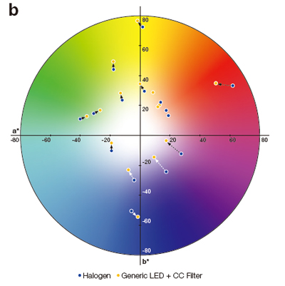
One way to compensate for this unwanted effect is to use a color correction (CC) filter. CC filters can absorb some of the light at wavelengths where the intensity is too high, creating a more balanced spectrum. The effect is shown in figure 4b; the colors with a CC filter are closer to halogen lamp illumination (shown by the shorter arrows), but there is still a considerable difference when compared with halogen lamp illumination.
Trust the Colors with Olympus True Color LED
|
Figure 4: Color Shifts
Chromaticity diagrams of halogen lamp vs LED (a) and halogen lamp vs LED + CC filter (b), show how colors shift when illuminated with a different light source.
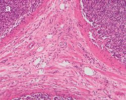
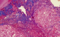
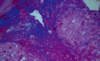
The practical effect of the color shift caused by a generic LED light source, both with and without a CC filter, is clearly visible when imaging a stained tissue section using the different light sources. Figure 5 shows a tissue section illuminated by the three different light sources mentioned before.
Figure 5: Stained Tissue Section
Differences between halogen lamp (a) and LED illumination (b) lead to tissue stainings shifting to blue, both through the oculars and on the screen. Adding a CC filter (c) mitigates the issue, but still gives a yellowish appearance.
UnparalleLED Color Rendering
To combine the benefits of LED – durable, bright and uniform illumination – with unparalleled color rendering, Olympus has developed the True Color LED light source. The name “True Color” refers to the fact that this new, redesigned light source specifically addresses the previously mentioned shortcomings and drastically improves the color rendering ability of LED equipped microscopes. Its spectrum (figure 6) has a much more uniform intensity and shows particular improvement in the cyan and red regions (arrows). The spectral improvements also mean that it closely matches halogen lamp illumination (blue line).
Trust the Colors with Olympus True Color LED
Figure 6: Robust in Red, Solid in Cyan |
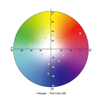
Figure 7: Spot-on |
The improved, more uniform spectrum has a considerable impact on the quality of light and this can be verified once again using a chromaticity diagram. Figure 7 shows that there are no significant differences between the color reproduction of halogen lamp and a True Color LED light source.
Case study: Comparing colors
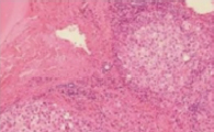
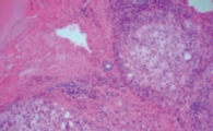
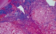
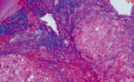
How does the Olympus True Color LED perform on real-world samples? To test this, the performance of the True Color LED was visually evaluated by direct comparison against other commercially available LED light sources on common histological stains: Hematoxylin and Eosin (H&E) and Azan Trichrome (figure 8).
The color rendering ability of the True Color LED not only closely mimics the reference halogen lamp light source but also stays constant regardless of the light intensity. This allows the operator to adjust the illumination without any distortions to the colors in a specimen.
|
|
|
|
|
|
|
|
Figure 8: Comparing LED Light Sources
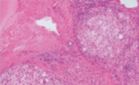
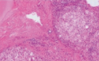
Both in H&E-stained (a-d) and in Azan-stained tissue sections (e-h) True Color LED illumination shows no discernable color shifts compared to halogen lamp whereas other commercially available LED sources show distinctive yellow shifts (c and g) or blue shifts (d and h).
Trust the Colors with Olympus True Color LED
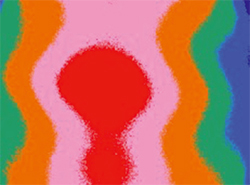
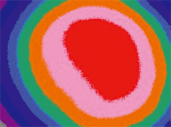
Olympus’ True Color LED chip has also been designed to produce a uniform illumination intensity over the entire field of view of the microscope. Uneven intensity can lead to artefacts when multiple images are stitched together and may require digital correction to produce high-quality images.
Figure 9 compares the intensity of different light sources across the field of view of a camera. The images show that as a result of the enhanced design of the True Color LED chip the illumination is greatly improved compared with the other types of illumination. This increased uniformity improves both the quality and the intensity of the colors in a specimen, thereby contributing to confidence in observations.
|
|
|
Figure 9: Intensity Measurement Across Field of View
Illumination intensity maps of a halogen lamp filament (a) a generic single-source LED (b) a True Color LED (c) across the camera’s field of view.
Summary
LED illumination holds great potential in clinical microscopy illumination due to its uniform intensity, high brightness, low energy consumption and long lifetime. However, many LED sources do not produce the same spectral uniformity as halogen lamp illumination and cause color shifts. Olympus’ market-leading True Color LED technology combines the benefits of LED technology with the capability to correctly show the sample colors thus providing clinical professionals, such as pathologists, with confidence when evaluating a stained sample.
Author
Flavio Giacobone
Clinical Vertical Market Specialist
Life Science EMEA
Scientific Solutions Division
OLYMPUS EUROPA SE & CO. KG
Sorry, this page is not
available in your country.
