November’s fan favorites were mainly very small samples with large personalities. From diatoms to Daphnia, let’s take a look!
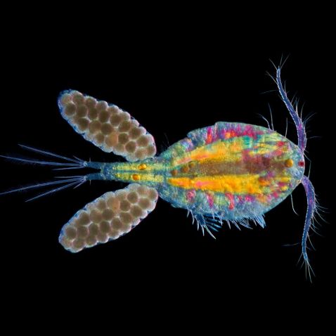
"I used darkfield illumination to produce a contrast between background and subject. Polarizing and wave plate retarder filters were then used to pick out the internal structures of the subject in color while oblique illumination provided some depth to the image."
Image captured with an Olympus BH-AAC condenser. Caption and Image courtesy of Anne Algar.

“Here is one of my favorite phytoplankton—the diatom Asterionella formosa. Forming star-like shapes with slender arms stretching out from the center, this colony was found in a water sample from a lake in the southern part of Sweden in late October (Vombsjön). During fall, cyanobacteria seem to decline in numbers as the water gets cooler. Instead, plenty of diatoms can be found. This time, Asterionella formosa and Fragilaria crotonensis were the two most dominant diatom species in the sample. Both are very beautiful.”
Caption and Image courtesy of Håkan Kvarnström.


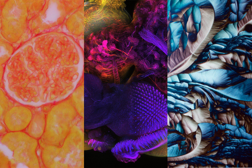
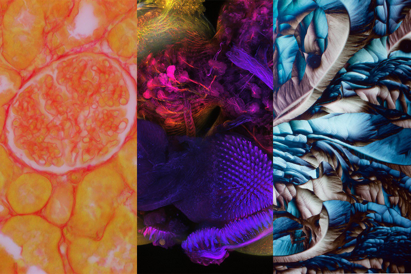
One of the best parts of the 2019 Global Image of the Year Award competition was seeing the variety of samples and imaging techniques submitted to the contest. Submit your best (3) images by January 10, 2021 for a chance to earn the 2020 Image of the Year title and win a SZX7 Stereo Microscope with DP27 Digital Camera!
Image 1: Drosophila (fruit fly) brains.
Image courtesy of Martin Hailstone. Captured on an Olympus FLUOVIEW FV3000.
Image 2: Bone fragments of sea cucumber.
Image courtesy of Yoshihiro Tamaru. Captured on an Olympus BH2.
Image 3: Amino acids.
Image courtesy of Karl Gaff. Captured on an Olympus BX51.
Image 4: Kidney section.
Image courtesy of Sara Al Marabeh. Captured on an Olympus BX53F with a DP72 camera.

A member of the family Daphnidae, the Simocephalus water flea engages in filter feeding while clinging to an object. Contrary to their name, they’re not actually fleas. Instead, they’re a type of small crustacean. And they’ve been around for a long time—they actually predate the Permian period, which ended about 250 million years ago!
Image courtesy of Anne Gleich.

“This ‘big’ 32-cell phytoplankton most likely is the species Pediastrum duplex. Around 100 micrometers across. Pediastrum are amazingly beautiful and come in many different shapes and forms, but most have the characteristic cogged wheel look. The ability to form colonies among phytoplankton acts as a morphological defense against grazers trying to eat the cells off a species. Larger phytoplankton normally experience lower mortality that smaller ones. Pediastrum form 2-dimensional plates of cells, as shown in the photograph. Some freshwater phytoplankton respond to infochemicals of various herbivores by forming larger colonies and thereby protecting themselves. It’s unclear if Pediastrum has that ability though, but fortunately it still often forms 32-cell colonies to the excitement of all microscopists.”
Caption and Image courtesy of Håkan Kvarnström.
To see more images like these, be sure to follow us on Instagram at @olympuslifescience!
Interested in sharing your own images?
Visit our image submission site or enter them into our 2019 Global Image of the Year contest.
Related Content
Brains and Blooms: Our Most Popular Microscope Images for October 2020
Revealing the Beauty of the Microscopic Scale—IOTY’s 2019 Asia Pacific Regional Winner
