While February is a short month, it was still full of beautiful images. From A to Z – that’s algae to zooplankton – here are the top images.
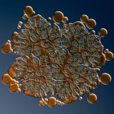
This month’s most popular image is the second in our new series with Håkan Kvarnström, "Exploring the Microscopic World with X Line Objectives."
Botryococcus braunii is a microalga notable for its copious oil production. In this image, you can see droplets of oil around the edges that were squeezed out of the specimen as water evaporated and the coverslip began to press down on the specimen.
To capture this focus stacked image, Håkan used the X Line 60X oil immersion objective (UPLXAPO60XO) with a numerical aperture (NA) of 1.42. The false-coloring effect was created by deliberate misalignment of the differential interference contrast (DIC) setup.
“The X Line 60X objective gave me the ability to resolve details that were hard to see with my other objectives. Its working distance is also considerably higher, so I can capture thicker specimens without losing sharpness and resolution.”
Keep your eyes peeled for next month's image to learn more about DIC imaging!
Captured with DIC using an X Line 60X objective. Image courtesy of Håkan Kvarnström.
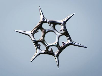
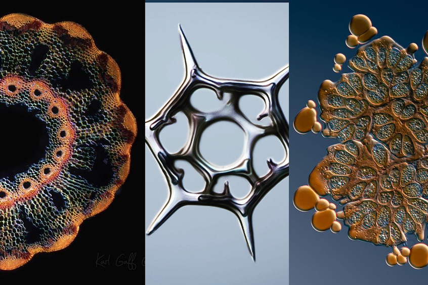
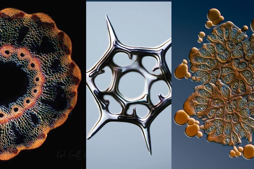
During their life cycle, Silicoflagellates produce a siliceous skeleton composed of a network of bars and spikes arranged to form an internal basket. Known as microfossils, these skeletons comprise 1–2% of the siliceous component of marine sediments. They evolved 120 million years ago during the Cretaceous, with the greatest abundance and diversity occurring during the Miocene, 25 million years ago.
Description and image courtesy of Håkan Kvarnström.
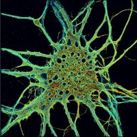


Bigger isn’t always better, especially in microscopy! This series of images shows how it is possible to reveal structures and dynamics at the nanoscale with Olympus optics and Abbelight’s barrier-breaking single-molecule localization microscopy (SMLM) technology.*1
Images left to right:
3D dSTORM image of cultured hippocampal neurons stained for β2-spectrin/alpha actinin and AF647-coupled secondary antibody. Sample and images courtesy of C. Leterrier, Neurocytolab, Marseille. Imaging performed on Abbelight SAFe360 mounted on an Olympus IX83 inverted miroscope, equipped with a 1.49 NA TIRF objective.
HeLa cells, microtubules (alphaTubulin antibody) + actin (phalloidin), 3D dSTORM. Image courtesy of Abbelight.*2
Mouse emrbyonal myoblasts, actin cytoskeleton stained with phalloidin-AF647and imaged via 3D dSTORM. Sample courtesy of Haniaa Segard, Team Bourdoulous, Institut Cochin, Paris; image by Abbelight.
HeLa cells, microtubules, alphaTubulin antibody, 3D dSTORM. Image courtesy of Abbelight.*2
*1 Abbelight partnership available only in the US and Canada.
*2 Although it became one of the most important cell lines in medical research, it’s imperative that we recognize Henrietta Lacks’ contribution to science happened without her consent. This injustice, while leading to key discoveries in immunology, infectious disease, and cancer, also raised important conversations about privacy, ethics, and consent in medicine.
To learn more about the life of Henrietta Lacks and her contribution to modern medicine, click here.
http://henriettalacksfoundation.org/
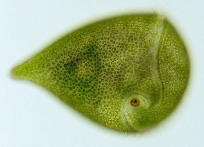
It’s not easy being green? It is for this protozoan! Known for its remarkable regenerative characteristics, it's able to grow back into a complete organism from a fragment as small as 1/100 its original size!
Image courtesy of Mr. Wim van Egmond.
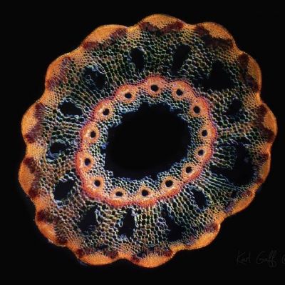
Did you know? The non-flowering evergreen perennial, horsetail dates back to Paleozoic times, some 350 million years ago. Like bamboo, it has vertical green stems with horizontal bands, and like ferns, its reproduction occurs through spores rather than seeds. However, horsetail is unrelated to bamboo or grass or ferns.
Specimen: Cross-section of the non-flowering evergreen perennial, Horsetail (Equisetum hyemale).
Image courtesy of Karl Gaff.
We always love our Instagram Takeovers when we hand the reigns over to microscopists and share the amazing work being done out in the world. This month, Gabrielle Corradino showed us her passion for plankton. Below are two of her zooplankton images.
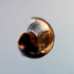

“What are zooplankton? Zooplankton are the animal-like plankton that have to hunt for their food within the water column. The water column is the area of water from the surface down to the sediment. During the daylight hours, zooplankton generally hang out in deeper waters to avoid predators. At night, these microscopic creatures venture up to the surface to feed, making this the largest migration on Earth. Check out @NASA images to see this migration from space!
Many species of zooplankton can be seen without a microscope and often look like tiny dots swarming around the water column. Common captures from a tow (a plankton net) include euphausiids (think krill!) and copepods. These are important grazers in the biological pump and can range from the poles to the warm tropics.”
To learn more about Gabrielle’s work, check out her interview.
Images and caption courtesy of Gabrielle Corradino.
To see more images like these, be sure to follow us on Instagram at @olympuslifescience!
Interested in sharing your own images? Visit our image submission site.
Related Content
Connecting with Curiosity—A Conversation with Dr. Gabrielle Corradino
From Algae to Art: Creating Stunning Microscope Artwork with X Line Optics
Dreaming of Spring—Our Most Popular Microscope Images for January 2021
