Evident LogoOlympus Logo

Parlez à nos experts

The Use of Multiplexing in Microscopy for Better Understanding the Skin Immune System in the Context of the Tissue

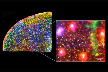
The skin is the first line of defense and the immune system’s biggest barrier for combating pathogens. Being able to accurately characterize and identify immune cell subtypes, tissues structures, and cell distribution in the skin under steady-state conditions provides a powerful tool for understanding the first immunological strategies and biological processes that occur in the presence of pathogens. In this webinar we will review technical aspects involved in the experimental process and explore how complementary imaging technologies might assist us to better understand the immune system.

The presentation is divided into three parts. First, an introduction of the Hugh Green Cytometry Centre will be presented and an overview of the histology and bioimaging technological platforms available. Second, the multiplexing methodology will be discussed, where several topics need to be considered for the design and development of a successful polychromatic panel for microscopy. Finally, preliminary results from a research project will be presented that constitutes part of a diploma program from The Royal Microscopical Society. The project focuses on the identification of immune cell types in the whole mount skin in relation to tissues structures (e.g., blood vessels and lymphatic network). It also centers on the immune cells’ distribution in the tissue as a first barrier of defense against pathogens.

Presenter: Alfonso J. Schmidt
Senior Staff Scientist, Malaghan Institute of Medical Research

Alfonso has a decade of experience working in a Shared Resource Lab (SRL) with a vast knowledge in histology, fluorescent microscopy, and image analysis. His work has been focused in maximizing the capabilities of the equipment available and in creating technical protocols and training modules for the scientific community. Currently, Alfonso oversees the Histology and Bioimaging Facility as part of the Hugh Green Cytometry Centre (HGCC) at the Malaghan Institute of Medical Research. Wellington, New Zealand.  

Produits associés

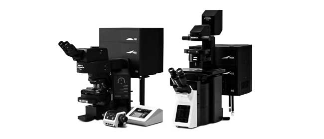
Microscope confocal à balayage laser

FV3000

  • Disponible en configuration galvanomètre seul (FV3000) ou hybride galvanomètre / résonant (FV3000RS)
  • Nouvelle détection TruSpectral haute efficacité et grande précision pour tous les canaux
  • Optimisé pour la prise d’images de cellules vivantes avec une sensibilité élevée et une faible phototoxicité
  • Statif inversé ou droit pour de nombreuses applications et types d’échantillons
Scanner de lames pour la recherche

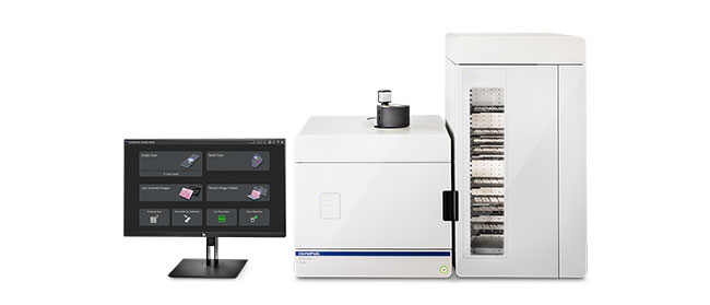
VS200

Le scanner de lames pour la recherche VS200 améliore la vitesse et l’efficacité des applications nécessitant une analyse quantitative d’un grand volume d’échantillons, notamment en recherche sur le cerveau, le cancer, les cellules souches et les médicaments. Prenez rapidement et facilement des images de lames en haute résolution, et analysez, partagez et archivez facilement vos données. Le système VS200 dispose de cinq modes d’imagerie différents : fond clair, fluorescence, fond noir, contraste de phase et polarisation simple. Il est possible de charger jusqu’à 210 lames à la fois pour une numérisation automatisée.

  • Imagerie de lames entières en haute résolution de 2X à 100X 
  • Compatible avec plusieurs tailles de lames et méthodes d’observation
  • Procédures simplifiées et efficaces, du fond clair au multiplexage de fluorescence
  • Possibilité unique de configuration du logiciel et de l’appareil pour des applications allant de l’acquisition manuelle des lames en fond clair à la reconnaissance par IA et à la numérisation entièrement automatisée

The Use of Multiplexing in Microscopy for Better Understanding the Skin Immune System in the Context of the Tissue

The skin is the first line of defense and the immune system’s biggest barrier for combating pathogens. Being able to accurately characterize and identify immune cell subtypes, tissues structures, and cell distribution in the skin under steady-state conditions provides a powerful tool for understanding the first immunological strategies and biological processes that occur in the presence of pathogens. In this webinar we will review technical aspects involved in the experimental process and explore how complementary imaging technologies might assist us to better understand the immune system.

The presentation is divided into three parts. First, an introduction of the Hugh Green Cytometry Centre will be presented and an overview of the histology and bioimaging technological platforms available. Second, the multiplexing methodology will be discussed, where several topics need to be considered for the design and development of a successful polychromatic panel for microscopy. Finally, preliminary results from a research project will be presented that constitutes part of a diploma program from The Royal Microscopical Society. The project focuses on the identification of immune cell types in the whole mount skin in relation to tissues structures (e.g., blood vessels and lymphatic network). It also centers on the immune cells’ distribution in the tissue as a first barrier of defense against pathogens.

Presenter: Alfonso J. Schmidt
Senior Staff Scientist, Malaghan Institute of Medical Research

Alfonso has a decade of experience working in a Shared Resource Lab (SRL) with a vast knowledge in histology, fluorescent microscopy, and image analysis. His work has been focused in maximizing the capabilities of the equipment available and in creating technical protocols and training modules for the scientific community. Currently, Alfonso oversees the Histology and Bioimaging Facility as part of the Hugh Green Cytometry Centre (HGCC) at the Malaghan Institute of Medical Research. Wellington, New Zealand.  

Produits associés

Microscope confocal à balayage laser

FV3000

  • Disponible en configuration galvanomètre seul (FV3000) ou hybride galvanomètre / résonant (FV3000RS)
  • Nouvelle détection TruSpectral haute efficacité et grande précision pour tous les canaux
  • Optimisé pour la prise d’images de cellules vivantes avec une sensibilité élevée et une faible phototoxicité
  • Statif inversé ou droit pour de nombreuses applications et types d’échantillons
Scanner de lames pour la recherche

VS200

Le scanner de lames pour la recherche VS200 améliore la vitesse et l’efficacité des applications nécessitant une analyse quantitative d’un grand volume d’échantillons, notamment en recherche sur le cerveau, le cancer, les cellules souches et les médicaments. Prenez rapidement et facilement des images de lames en haute résolution, et analysez, partagez et archivez facilement vos données. Le système VS200 dispose de cinq modes d’imagerie différents : fond clair, fluorescence, fond noir, contraste de phase et polarisation simple. Il est possible de charger jusqu’à 210 lames à la fois pour une numérisation automatisée.

  • Imagerie de lames entières en haute résolution de 2X à 100X 
  • Compatible avec plusieurs tailles de lames et méthodes d’observation
  • Procédures simplifiées et efficaces, du fond clair au multiplexage de fluorescence
  • Possibilité unique de configuration du logiciel et de l’appareil pour des applications allant de l’acquisition manuelle des lames en fond clair à la reconnaissance par IA et à la numérisation entièrement automatisée
Experts
Alfonso J. Schmidt
Senior Staff Scientist
Malaghan Institute of Medical Research

Alfonso has a decade of experience working in a shared resource lab (SRL) with a vast knowledge in histology, fluorescent microscopy, and image analysis. His work has been focused in maximizing the capabilities of the equipment available and in creating technical protocols and training modules for the scientific community. Currently, Alfonso oversees the Histology and Bioimaging Facility as part of the Hugh Green Cytometry Centre (HGCC) at the Malaghan Institute of Medical Research in Wellington, New Zealand.

The Use of Multiplexing in Microscopy for Better Understanding the Skin Immune System in the Context of the Tissuenov. 25 2024
Sorry, this page is not available in your country
Ask the Expert Sign-up

By clicking subscribe you are agreeing to our privacy policy which can be found here.

Sorry, this page is not
available in your country.

Sorry, this page is not available in your country