Not Available in Your Country
Sorry, this page is not
available in your country.
Overview
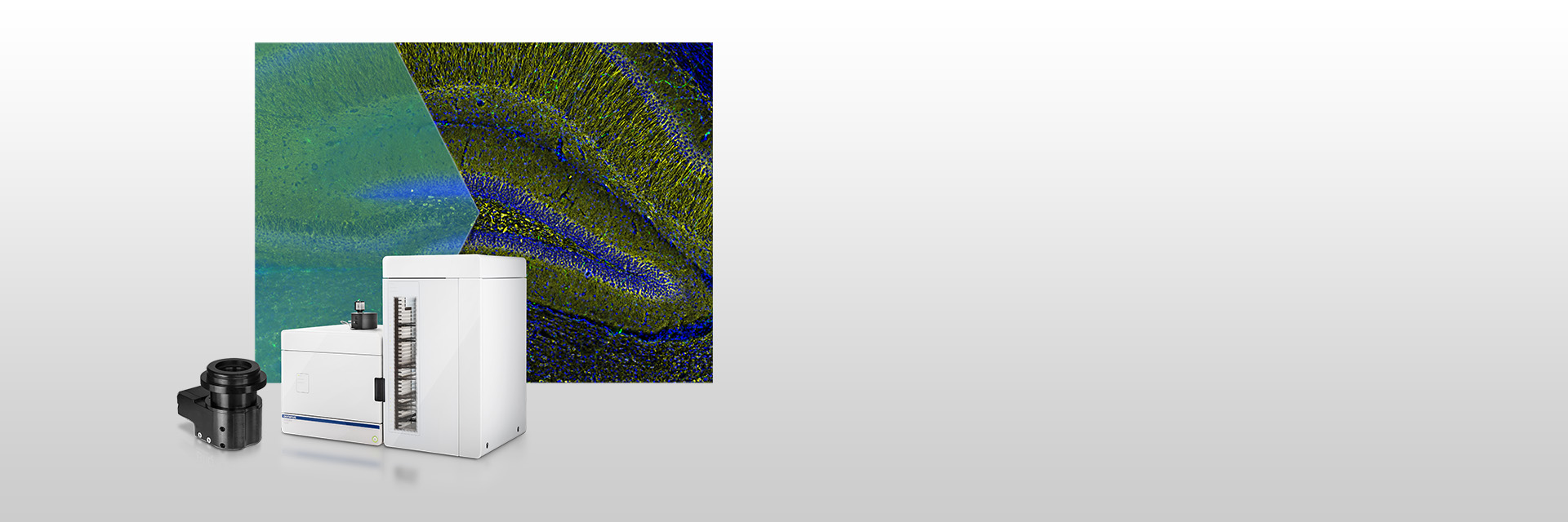 | High-Contrast Optical Sectioning ImagesThe SILA (Speckle Illumination Acquisition) optical sectioning device easily integrates into your VS200 system and software. Speckles are used to obtain high-contrast images, removing out-of-focus light to deliver sharp images, especially from thick samples. The image is computed during the scan, and there’s no need for post-processing, so the device has a minimal impact on acquisition speed. |
|---|
Optical Sectioning TechnologyThe VS200 SILA device—developed by Bliq Photonics—is a camera-based optical sectioning technology. This optional feature for VS200 scanners delivers real-time, blur-free scans of thick samples, so you can achieve widefield and section imaging in a single system with up to 6 laser lines. | 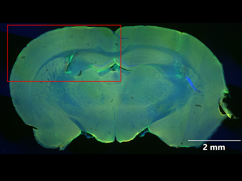 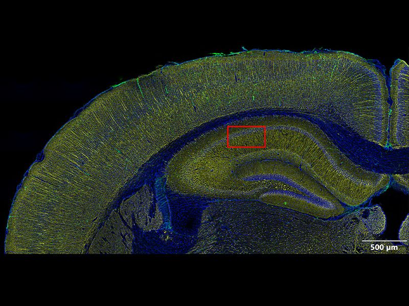  |
| Easy to UseThe SILA device is easy to use with samples of any thickness and independent of the magnification. No special calibration is required, and it’s compatible with a variety of existing Evident objectives. Just insert the slide manually or using the loader, load the software, and acquire the images with the ‘SILA’ option enabled. |
|---|
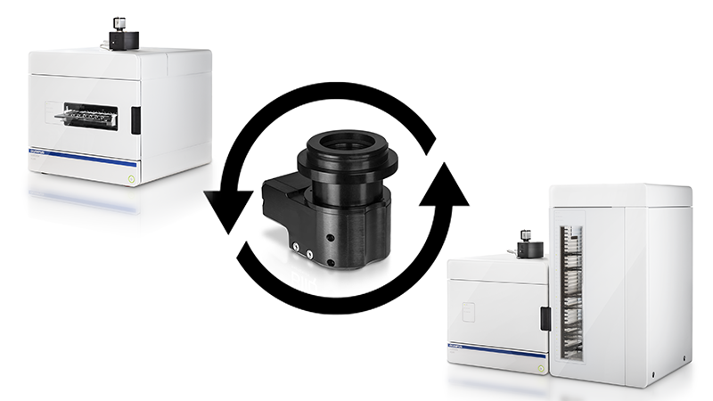
Compact and Easy to InstallThe SILA module is compact. It can be used with any manual or loader VS200 system, and existing VS200 scanners can be upgraded on site. | Related Videos |
 Widefield (left) and SILA (right) images of a whole planarian flatworm Schmidtea mediterranea in 20x, showing the intestines. Blue: DAPI. Green: inner intestine cells; Red: outer intestine cells. Samples provided by Amrutha Palavalli, Department for Tissue Dynamics and Regeneration, Max Planck Institute for Multidisciplinary Sciences, Goettingen, Germany. | Versatile for Many ApplicationsThe device works with most sample types, including cleared and fixed cells/tissues. Using optical sectioning, it is possible to image thick samples well beyond the limit of a regular widefield microscope and at any magnification. |
|---|
Need assistance? |
Applied Technologies
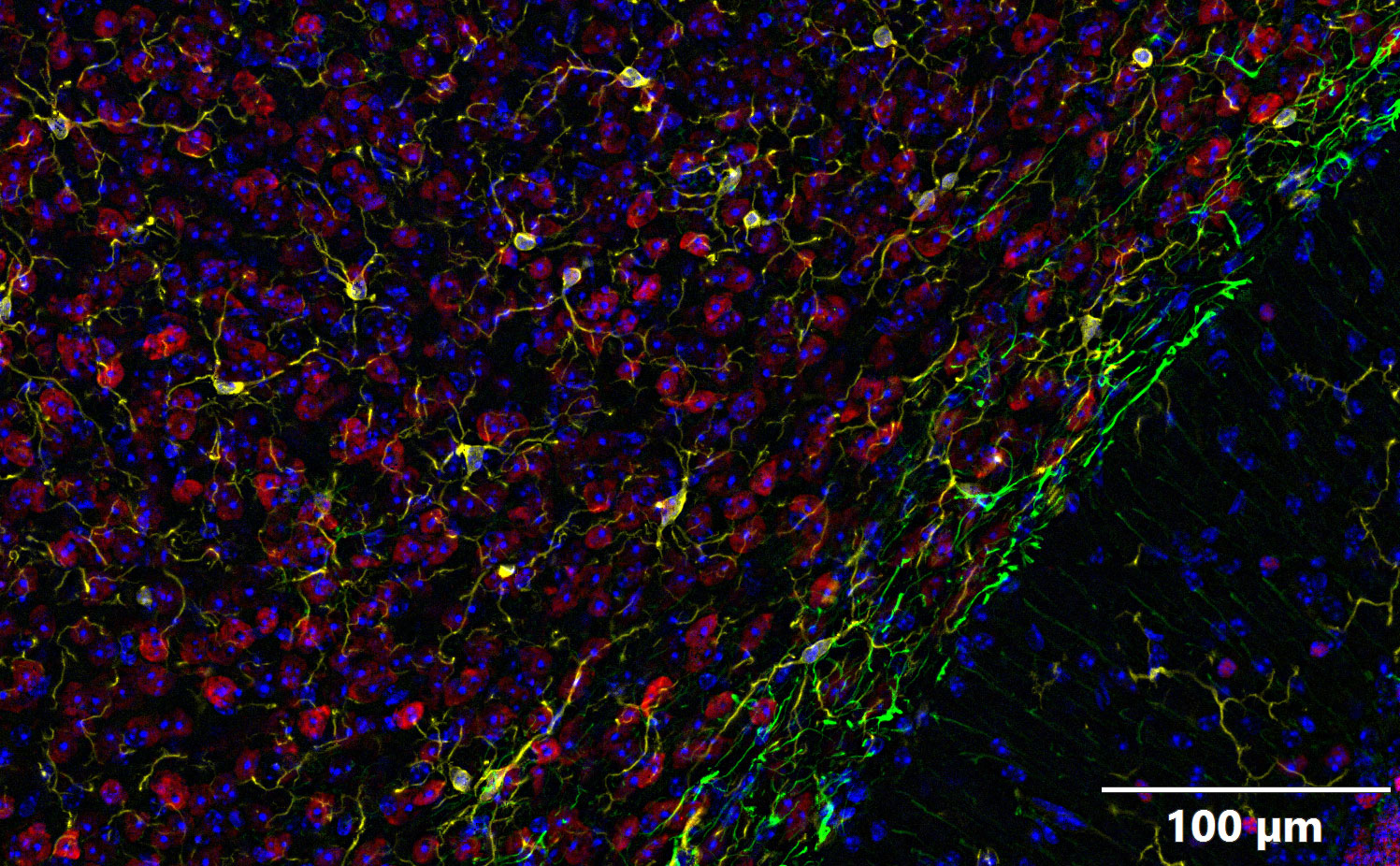
Sharp Images from Deep SamplesThe technology offers a high penetration depth. Since the speckles remain sharp at depth, you retain the same optical sectioning capabilities as you go deeper. | Related VideosOverview acquired at 4x, detailed scan is an extended focal image (EFI) of a 19um Z series acquired at 20x . Blue: DAPI, Green: GFAP (astrocytes), Yellow: IbaI (microglia). Red: NeuN (neurons). Samples provided by Institute of Experimental Neuroregeneration, Spinal Cord Injury and Tissue Regeneration Center Salzburg (SCI-TReCS), Paracelsus Medical University, Austria. |
Easy to AdjustThe SILA optical sectioning device scans over large field of views in a short amount of time. |
|
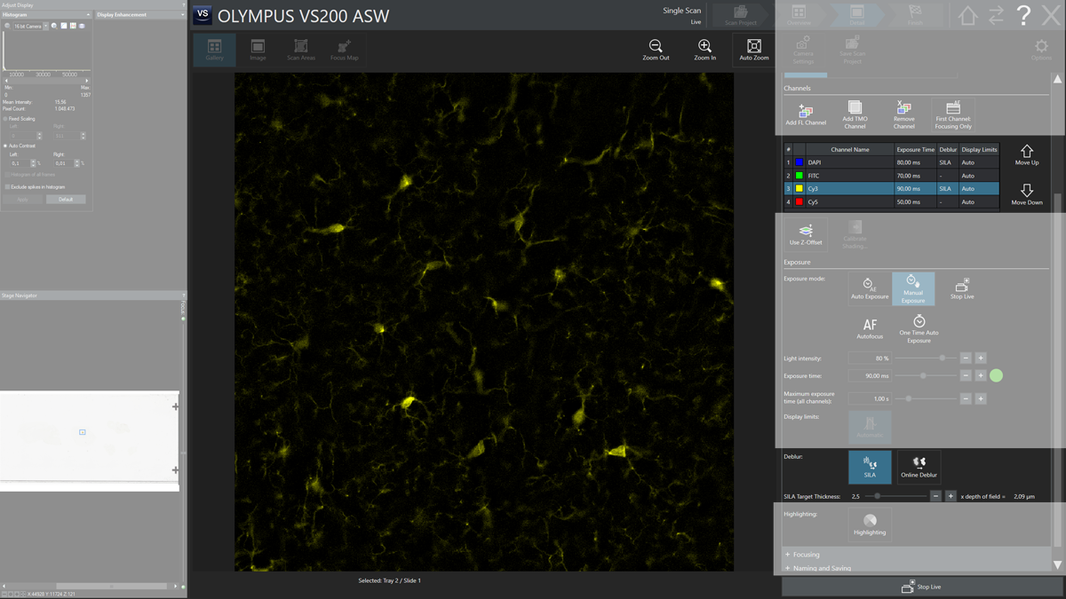
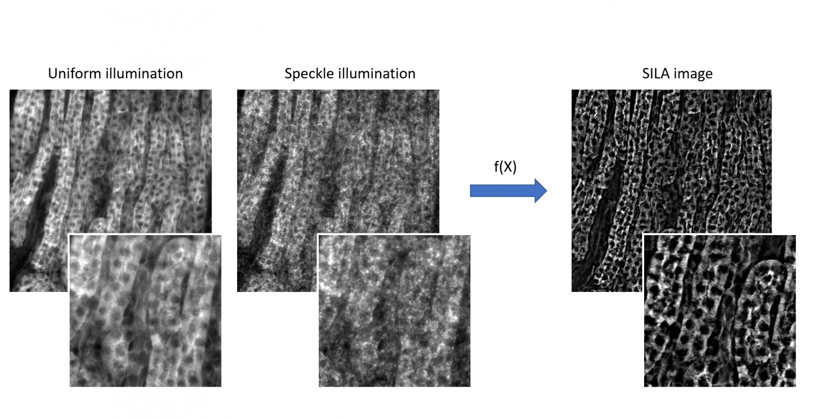
Fast Processing with the Ability to Visualize Optically Sectioned Images in Live ViewThe SILA optical sectioning device rapidly removes out-of-focus elements using two illuminated images that are mathematically processed. Based on HiLo microscopy technology, the image is obtained using a combination of hardware and software. The SILA calculations run on a GPU during image acquisition, so it is also possible to preview the optical sectioning capabilities directly on the live image. Use one parameter to remove out-of-focus light above and below the focus plane, similar to adjusting a confocal pinhole.
|
|---|
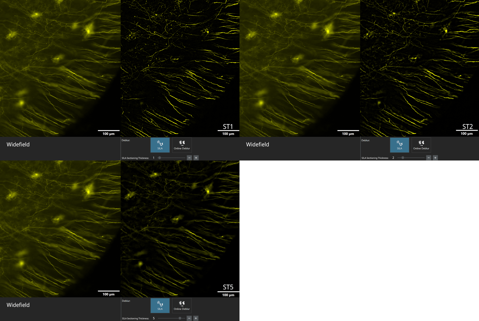
Simple for Anyone to UseOnly one parameter needs to be adjusted to obtain the sectioned images: the sectioning thickness. There’s no need to post-process the images like in deconvolution and no need to calculate the point spread function (PSF) of the objects. | Related Videos |
| Easily Upgrade Your Existing VS200 Slide ScannerThe SILA module can be ordered as an upgrade kit and easily installed by our service personnel to any VS200 base unit or loader system, making it possible to obtain optically sectioned images without buying a new system. |
|---|
Need assistance? |
Applications
Works in a Range of ApplicationsUse the SILA optical sectioning device to image cleared, fixed samples of different thicknesses. It is especially useful for neurobiology, botany, organoid research, entomology, developmental biology, and tissue regeneration.
|
SILA image of a 40um mouse brain sagittal section. Overview acquired at 4x, detailed scan is an extended focal image (EFI) of a 19um Z series acquired at 20x . Blue: DAPI, Green: GFAP (astrocytes), Yellow: IbaI (microglia). Red: NeuN (neurons). Samples provided by Institute of Experimental Neuroregeneration, Spinal Cord Injury and Tissue Regeneration Center Salzburg (SCI-TReCS), Paracelsus Medical University, Austria. | Flexible Fixed Cell ImagingAcquire 2D and 3D images: (live SILA image to set up the conditions), Z-stacks, stitching, and EFI. Multichannel acquisition: can be used with up to 4 channels (405, 488, 561, and 638 nm excitation wavelengths). |
|---|
Optimized for Thick SamplesObtain a high penetration depth as the speckles remain sharp the deeper you go. | Related Videos |
Need assistance? |
Specifications
| Compatible System | VS200 SLIDEVIEW Slide Scanner with Fluorescence | |
|---|---|---|
| Intended Specimen | Observable Specimen | Glass slide with cover glass |
| Thickness of Cover Glass | 0.12–0.17 mm | |
| Thickness of Specimen | Up to 0.3 mm | |
| Imaging | Observation Methods | Fluorescence—optical sectioning with speckle illumination |
| Magnification | 4x, 10x, 20x, 40x, 60x, and 100x (incl. selected oil immersion and silicon oil immersion objectives) | |
| Sectioning Thickness | 1 to 10 x depth of field of the objective in use | |
| Exposure | Minimum: 50 ms | |
| Scan Time |
Approx 14 minutes
(20x objective scan area 15 mm × 15 mm, 4 channels, 50 ms exposure each) | |
| Light Source | Wavelengths | Range of 375–785 nm supported; compatible filters required. |
| Optical Fiber | Single mode fiber (connector: FC/APC) | |
| Laser combiner |
Laser power:
375 nm: 70 mW 730 nm: 50 mW Other wavelengths: 100 mW AC adapter: 15 VDC, 7A | |
| Software Solution | ASW solution license | SILA acquisition (VS20-SILA) |
| SILA Hardware |
Dimensions
(H × W × D) |
72.0 mm × 98.7 mm × 88.5 mm (2.8 in. × 3.9 in. × 3.5 in.)
Weight: 520 g (1.1 lb) |
| Operating Environment |
Temperature: 12–28 °C (53.6–82.4 °F)
Humidity: up to 80% (31 °C or 87.8 °F) Altitude: 2000 m (6561.7 ft) | |
| Power | 5 VDC, 0.5 A | |
| TTL control Input |
SMA connector
Threshold: 3.0 V | |
