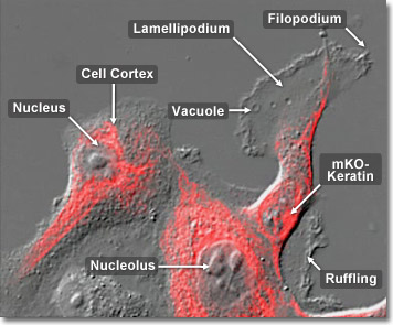
Cytokeratins are a class of intermediate filaments found in the cytoplasmic cytoskeleton of epithelial tissues. There are two distinct types of cytokeratins: the low molecular weight, acidic type I cytokeratins and the high molecular weight, basic or neutral type II cytokeratins. Cytokeratins are usually found in pairs composed of a single type I and a single type II. Because the cytokeratin distribution profile tends to remain constant when epithelial tissue undergoes malignant transformation, it is a tool of immense value in tumor diagnosis and surgical pathology. In the digital video presented above, rabbit kidney epithelial cells (Rk-13 line) are expressing a monomeric Kusabira Orange (mKO) fluorescent protein fusion to human cytokeratin.
Video 1 - Run Time: 58 Seconds
Video 2 - Run Time: 30 Seconds
Video 3 - Run Time: 17 Seconds
Video 4 - Run Time: 34 Seconds
Video 5 - Run Time: 48 Seconds
Video 6 - Run Time: 57 Seconds
Video 7 - Run Time: 35 Seconds
Video 8 - Run Time: 34 Seconds
Video 9 - Run Time: 28 Seconds
Video 10 - Run Time: 57 Seconds
Video 11 - Run Time: 30 Seconds
Intermediate filaments are one of three types of cytoskeletal elements. The other two are thin filaments (filamentous actin) and microtubules. Frequently the three components work together to enhance structural integrity, cell shape, as well as cell and organelle motility. Intermediate filaments tend to be both stable, and durable, ranging in diameter from 8 to 10 nanometers, a length considered intermediate in size compared with thin filaments and microtubules. They are prominent in cells that withstand mechanical stress and are the most insoluble part of the cell. In the digital video presented above, rabbit kidney epithelial cells (Rk-13 line) are expressing a monomeric Kusabira Orange (mKO) fluorescent protein fusion to human cytokeratin.

The subunits of intermediate filaments are elongated, not globular, and are associated in an anti-polar orientation. As a result, the overall filament has no polarity, and therefore motor proteins are unable to move along intermediate filaments. Found only in complex multicellular organisms, intermediate filaments are encoded by a large number of different genes and can be grouped into families based on their amino acid sequences. Cells in different tissues of the body express one or another of these genes at different times. In general, intermediate filaments serve as structural elements, helping cells maintain their shape and integrity. In the digital video presented above, rabbit kidney epithelial cells (Rk-13 line) are expressing a monomeric Kusabira Orange (mKO) fluorescent protein fusion to human cytokeratin.