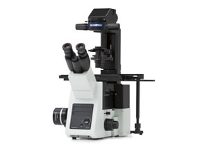
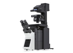
High-Resolution Objectives for TIRFA high numerical aperture (NA) is important for TIRF microscopy. As a pioneer in TIRF microscopy, we offer a diverse lineup of objectives with a high NA, ranging from 1.45 to the world’s highest NA of 1.7*1and from 60x to 150x magnification. Modern observation methods, like super resolution and capturing images over a large field of view with an sCMOS camera, demand the highest quality objectives. That’s why we developed advanced objective manufacturing technology. This advanced polishing technology enabled us to create the world’s first plan-corrected apochromat objectives with an NA of 1.5*2. These objectives deliver uniform image quality, even over a large field of view, and are ideal for TIRF imaging. *1 As of Nov, 2018. According to Olympus research. | 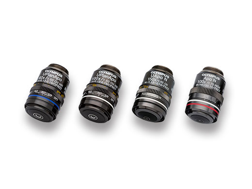 |
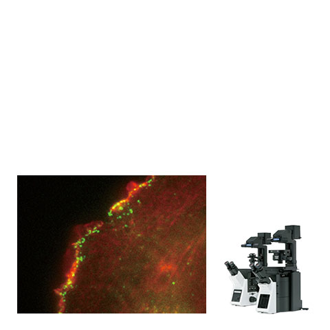
Time–Lapse TIRF Imaging of Polymerization and Depolymerization Between a Plasma Membrane and FBP17 Membrane–Bending Protein Part 1.The penetration depth was adjusted to get a high signal-to-noise ratio with a high numerical aperture. |
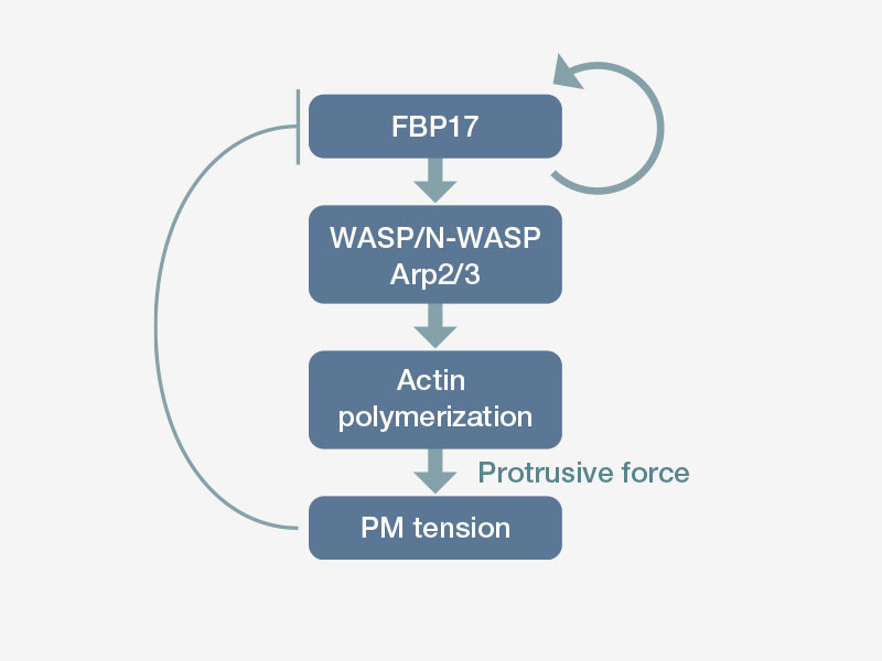
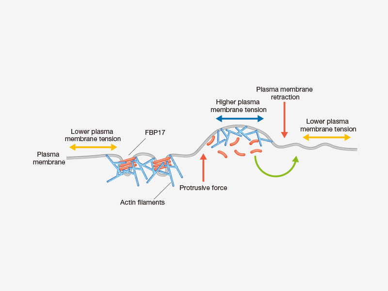
Videos asociados | Time–Lapse TIRF Imaging of Polymerization and Depolymerization Between a Plasma Membrane and FBP17 Membrane–Bending Protein Part 2.Researchers examined whether the recruitment of FBP17 to the plasma membrane is dependent on a transient reduction of membrane tension caused by myosin-based contraction force. FBP17 acutely disappeared from the cell edge after treatment with the myosin inhibitor blebbistatin (175 sec). The images were acquired using the UAPON100XOTIRF.*4 Image data courtesy of Kazuya Tsujita, Ph.D., Toshiki Itoh, Ph.D., Biosignal Research Center, Organization of Advanced Science and Technology, Kobe University Reference: Nat Cell Biol. 2015 Jun; 17 (6): 749–58. doi: 10.1038/ ncb3162. |
|---|
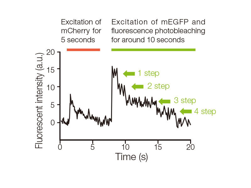
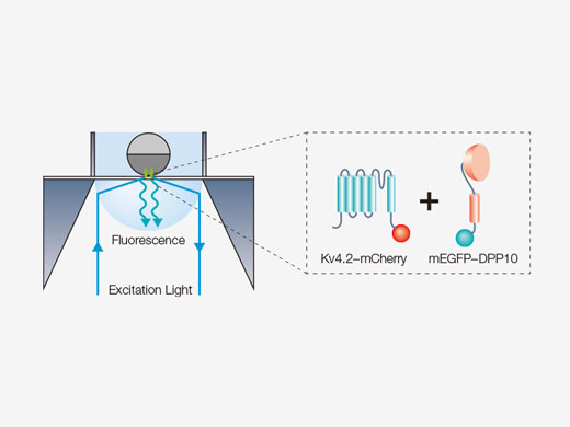
Single–Molecule Fluorescence Imaging to Count the Subunits of a Transmembrane Ion Channel Complex (APON100XHOTIRF) Part1.The objective enables single-molecule TIRF imaging with higher resolution and brighter images thanks to a numerical aperture of 1.70. The subunit counting*5 technique was used to analyze the number of accessory dipeptidyl peptidase-like protein 10 (DPP10) molecules, which bind to the transmembrane ion channel Kv4.2, in one Kv4.2–DPP10 complex. The APON100XHOTIRF objective’s high NA enables researchers to measure fluorescence intensity change caused by photobleaching of a single molecule. This study*6 revealed that a maximum of 4 molecules of DPP10 subunits form a complex with the ion channel Kv4.2. *5 Ulbrich, MH, and Isacoff EY. “Subunit counting in membrane–bound proteins.” Nature Methods, 4 (2007): 319–321. |
|
Single–Molecule Fluorescence Imaging to Count the Subunits of a Transmembrane Ion Channel Complex (APON100XHOTIRF) Part2.Localization of Kv4.2–mCherry is visualized by the excitation of mCherry in the first 5 seconds, followed by the excitation of mEGFP in the next 10 seconds to visualize its localization and continuous fluorescence photobleaching. Spots with photobleaching of mEGFP in a maximum of 4 steps were found by graphing the change in fluorescence intensity at each spot where two colors of fluorescent molecules colocalized (indicated by the white arrow head). Therefore, it was found that a maximum of 4 molecules of mEGFP–DPP10 were bound in the Kv4.2 ion channel complexes. Image data courtesy of; Masahiro Kitazawa, Ph.D., Yoshihiro Kubo, M.D.,Ph.D., Koichi Nakajo*, Ph.D. , Division of Biophysics and Neurobiology, Department of Molecular Physiology, National Institute for Physiological Sciences*Present address: Department of Physiology, Osaka Medical College |
|---|
Selection Guide of TIRF Objectives
|
Working Distance
(mm) | Magnification | Objective Field Number*3 | Numerical Aperture | Cover Glass | Immersion | |
|---|---|---|---|---|---|---|
| UPLAPO60XOHR | 0.11 | 60X | 22 | 1.50 | 0.13-0.19 | Oil |
| UPLAPO100XOHR | 0.12 | 100X | 22 | 1.50 | 0.13-0.19 | Oil |
| APON100XHOTIRF | 0.08 | 100X | 22 | 1.70 | 0.15 | Special Oil |
| UAPON150XOTIRF | 0.08 | 150X | 22 | 1.45 | 0.13-0.19 | Oil |
*3 Maximum field number observable through eyepiec
Sorry, this page is not
available in your country.
Sorry, this page is not
available in your country.

.jpg?rev=1AF2)
