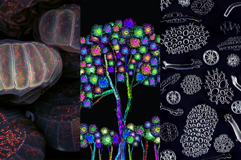
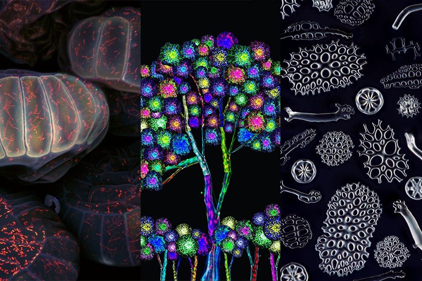
This month’s top images include some great examples of how scientists can manipulate their subjects via techniques such as staining, sample placement, and image stacking to make even more beautiful works of sciart.
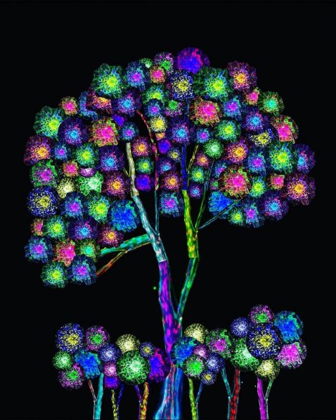
Appropriately titled "Tree of Nerve," this microscope image is not a plant or flower but instead a beautiful collection of nerves. The flowers are made from dorsal root ganglion cluster and schwann cells explant cultures, while the branches and trunks are made from sciatic nerves stained for schwann cells and neurofilament. A true example of the art of science!
Image courtesy of Diara Santiago Gonzalez. Captured with an Olympus DSU.
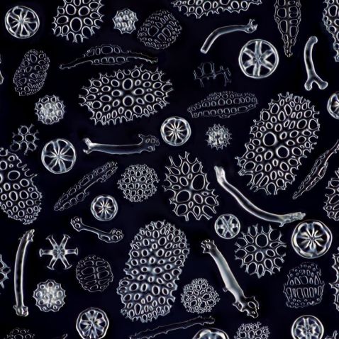
While named after their resemblance to the vegetables, sea cucumbers are marine animals. While their leathery skin and elongated body might not make them the most striking of nature’s creatures, they serve a useful role in the marine ecosystem as they help recycle nutrients, breaking down detritus and other organic matter. This is a close-up look at the various types of bone fragments of sea cucumber.
Image courtesy of Yoshihiro Tamaru. Captured on an Olympus BH2 microscope at 10x magnification.

Did you know? Ferns do not grow from seeds and flowers but, in fact, reproduce from spores that form on the underside of the leaves, seen here. There are more than 10,000 known extant species of fern.
Captured on a FLUOVIEW FV3000 microscope.

“This is close-up look at a Nauplius larvae of a Cyclops Copepod. Around 0.1 mm in size, they are predators and very efficient hunters. They use their strong legs (antennas) to jump over their prey and kill them within 50 milliseconds. They are extremely fast and can move at a speed up to 100 body lengths per second during an attack or when trying to escape bigger predators. Later in life, these larvae develop into adult Cyclops (Copepods). Check out the “muscles” of the antennas! No wonder it can move so fast. As all true Cyclops, they have a single red eye (photo receptor) in the front center of the body.”
Caption and Image courtesy of Håkan Kvarnström. Captured on an Olympus BX51 microscope with a 10X X Line objective and DIC.

Female daphnia, or water fleas, produce a brood of diploid eggs that will stay in the brood pouch for up to three days after hatching. These broods can contain as few as 1–2 eggs in smaller species, but as many as 100 or more in larger species. This darkfield illumination image of a female is made up of 69 total images.
Image courtesy of Christopher Algar. Captured on an Olympus BHB microscope.
Videos asociados
Bonus video! Because tardigrades are terrific!
“A tardigrade is attempting to walk along my microscope slide. There are over 1,300 known species of tardigrade. I saw that someone posted one the other day that looked like a gecko!!! Does anyone remember who posted that?? I like how the stylets coming from the mouth shine a pretty color in the polarized Ret. light. The part that looks kinda like a brain is actually the pharynx; it still does have a brain, but it’s hard to see and located more near the eyes.”
Image and caption courtesy of Logan Upclose.
To see more images like these, be sure to follow us on Instagram at @olympuslifescience!
Interested in sharing your own images?
Visit our image submission site.
Related Content
Barrier-Breaking and Now Edison Award-Winning Objectives
On the Road to a Breakthrough—Trailblazing Bioimaging Is Transforming Cancer Research
How to Clean and Sterilize Your Microscope