Judges 2008

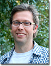
 | Mr. Wim van Egmond - A winner of numerous awards in the past few years for his stunning photography through the microscope, Wim van Egmond is a freelance photographer based in Rotterdam, the Netherlands. Mr. van Egmond specializes in microscopy of small water-borne creatures using a variety of advanced contrast-enhancing techniques, such as phase contrast, darkfield, and differential interference contrast. He is considered a world-renown expert in stereo imaging for optical microscopy and has produced a large collection of exhibitions, articles, and image galleries that are posted in numerous spots on the web. Mr. van Egmond is also a custom web designer who has completed several commercial, artistic, and educational sites, including the Smallest Page on the Web and the Virtual Pond Dip. |
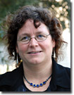
 | Dr. Claire Brown - An expert on fluorescence correlation spectroscopy (FCS) and laser scanning confocal microscopy, Dr. Brown is the Imaging Facility Director for the McGill University Life Sciences Complex in Montreal, Quebec, Canada. Dr. Brown has written numerous research articles in FCS as well as educational reviews on the basics of confocal and fluorescence microscopy. Among the techniques offered by Dr. Brown's facility are laser scanning confocal, total internal reflection (TIRF), live-cell imaging, laser micro-dissection, resonance energy transfer (FRET), fluorescence recovery after photobleaching (FRAP), deconvolution, and image correlation spectroscopy. Perhaps the best known faucet of Dr. Brown's teaching efforts is a popular poster entitled: Fluorescence Microscopy - Avoiding the Pitfalls, which was published (and is available as a cost-free download) with an accompanying article in the Journal of Cell Science in 2007. |
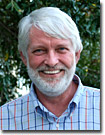
 | Dr. John M. Murray - Professor Murray is a faculty member in the Cell and Developmental Biology Department at the University of Pennsylvania School of Medicine. In addition to his faculty duties and research, Dr. Murray is also a avid participant in workshops designed to teach advanced microscopy techniques at the Cold Spring Harbor Laboratory in Long Island, New York. Dr. Murray's research focuses on the replication and assembly of daughter cells of the human pathogen Toxoplasma gondii, which infects a third of the population. In addition to his expertise in all forms of optical microscopy and live-cell imaging, Dr. Murray is also heavily involved in electron microscopy to solve biological problems. He is the author of numerous scientific and educational publications, including an excellent review article on laser scanning, multiphoton, and spectral imaging microscopy in the popular Live-Cell Imaging handbook published by the Cold Spring Harbor Press. |
 | Dr. Douglas B. Murphy - Professor Murphy is the Director of the optical and electron microscopy laboratory at the Howard Hughes Medical Institute in Janelia Farm, Virginia. This resource is a core facility that provides equipment and services in fluorescence, confocal, and electron microscopy to laboratories throughout the research campus. Dr. Murphy's research interests include the dependence of microtubules in organelle transport and related microtubule behavior, including polymer annealing and the mechanism of binding microtubule-associated protein (MAP-2) on microtubule surfaces. He has also explored the mechanical and conformational requirements of the motor protein, kinesin. The core facility specializes in advanced fluorescence techniques (FRET, FRAP, TIRFM), confocal, multiphoton, and time-lapse in multiple fluorescence channels. Dr. Murphy has been a significant contributor to review articles and Java tutorials on the Olympus optical microscopy educational websites. |
Not Available in Your Country
Sorry, this page is not
available in your country.