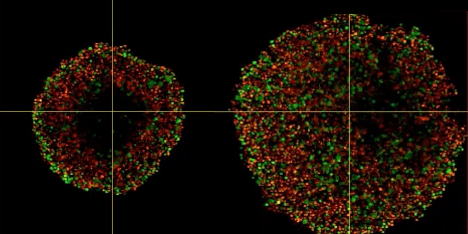
Phenotypic tumour heterogeneity arising due to differentially cycling cell populations has been implicated in increased therapy resistance. This phenomenon cannot be assessed in adherent cell culture, where microenvironmental conditions are homogeneous. Thus, we utilise melanoma spheroids to model the 3D tumour microenvironment including the extracellular matrix (ECM) and study spheroid structure, necrotic region, individual cell arrangement within and gene expression patterns. We achieve this by exploiting the fluorescence ubiquitination cell cycle indicator (FUCCI) system to monitor cell cycle stages as a surrogate marker for phenotypic tumour heterogeneity, tissue clearing and confocal microscopy using FV3000.
Investigating Spheroid Architecture Using the FV3000 Confocal MicroscopePhenotypic tumour heterogeneity arising due to differentially cycling cell populations has been implicated in increased therapy resistance. This phenomenon cannot be assessed in adherent cell culture, where microenvironmental conditions are homogeneous. Thus, we utilise melanoma spheroids to model the 3D tumour microenvironment including the extracellular matrix (ECM) and study spheroid structure, necrotic region, individual cell arrangement within and gene expression patterns. We achieve this by exploiting the fluorescence ubiquitination cell cycle indicator (FUCCI) system to monitor cell cycle stages as a surrogate marker for phenotypic tumour heterogeneity, tissue clearing and confocal microscopy using FV3000. | |
Productos relacionadosMicroscopio confocal de escaneo láser FV3000
| |
Investigating Spheroid Architecture Using the FV3000 Confocal Microscope
Videos asociados
Productos relacionados
FV3000
- Disponible únicamente con las configuraciones de escaneo galvanométrico (microscopio de escaneo FV3000) o híbrida galvanométrica-resonante (FV3000RS)
- Nueva detección altamente eficaz y precisa en todos los canales mediante la tecnología TruSpectral
- Optimizada para el tratamiento de imágenes de células vivas proporcionando alta sensibilidad y baja fototoxicidad

