Université de Montréal
| |||||
| “The partnership between Evident and our School of Optometry as well as our CIRCA Research Center will contribute to enhance the quality and impact of research as well as the training of students by facilitating access to advanced equipment and savoir-faire. The Université de Montréal is very proud of this new collaboration.”―Professor Yves Joanette, Deputy Vice-Principal of Research at UdeM |
In the Spotlight | |
Discovery Centre Inauguration   | Ribbon cutting (left to right):
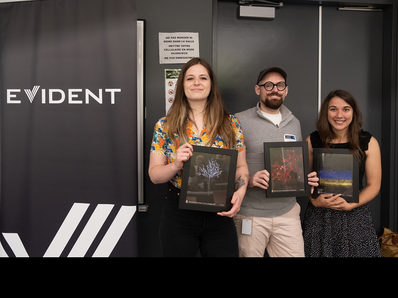
Image contest winners (left to right):
|
Image contest winners | ||
First place: Chloé Savard This microscopic tree looks like a plant but moves like an animal, but what is it exactly? It’s neither a plant nor an animal, but a colony of single cell organisms called ciliates from the Carchesium polypinum species. This microbe can be found around the globe, mainly in freshwater ponds and lakes but is also used in sewage treatment. Carchesium possess thousands of tiny cilia that create a water vortex drawing food particles toward the colony and each single cell. They usually eat bacteria, phytoplankton and debris that are floating around them. The stalks of these creatures have the ability to contract like springs in a matter of 10-20 mm per second and becomes 20 to 40% shorter as their bell becomes spherical! The bell of these ciliates can reach a speed up to 60-90 mm per second which corresponds to 1200 body length per second and makes them one the fastest living organisms! Captured using darkfield imaging. |
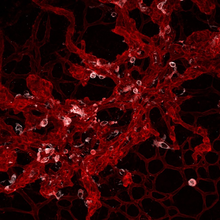
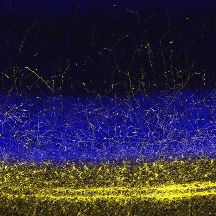
Second place: Sergio Crespo-Garcia Mononuclear phagocytes populating a site of pathological angiogenesis in the ischemic mouse retina. Captured on an Olympus FLUOVIEW FV1000 confocal microscope. | Third place: Catarina Sofia Micaelo Fernandes Au-dessus des étoiles / Above the stars ; Axones exprimant le CB1R (en bleu) au-dessus des "étoiles" gliales exprimant la GFAP (en jaune) Cortex visuel du singe vervet, transition cortex/substance blanche. Captured on an Olympus FLUOVIEW FV3000 confocal microscope. |
Systems at Université de Montréal | |
FLUOVIEW™ FV3000 upright confocal laser scanning microscope
Enabling the observation and recording of fast phenomena and live physiological events, this hybrid laser scanning unit uses a galvanometer scanner for precision scanning and a resonant scanner for high-speed imaging with a with a large field of view. | PhaseView® Alpha3 light sheet microscope
Offering researchers a flexible, high-performing light sheet solution, the Alpha3 system enables high-speed, intelligent optical focus sweeping for uniform, artifact-free, multidimensional imaging across the entire field of view. |
SLIDEVIEW™ VS200 slide scanner
Capturing high-resolution digital slide images for quantitative analysis, the VS200 system with 210-capacity slide loader enables researchers to easily analyze, share, and archive their data to fully leverage the information their slides have to offer. | |
Not Available in Your Country
Sorry, this page is not
available in your country.


