CX41 Upright Microscope
Discontinued Products

This product has been discontinued, check out our current product
The CX41 biological microscope sets the standard for its class in both basic and system performance. It provides high image clarity in a variety of observation methods, including bright field, phase contrast and fluorescence. This cost-efficient microscope is suitable for routine observation and has an ergonomic design for comfortable long-term use. The renowned UIS2 plan-corrected objectives result in outstanding flat images. Olympus digital camera combinded with software packages to allow efficient documentation, analysis and reporting.
Not Available in Your Country
Sorry, this page is not
available in your country.
Features
Advanced Optical and System Performance
The CX41 is the cost-efficient microscope system for all inspections and training applications in the field of biology and medicine. Due to its outstanding performance in combination with the renowned UIS2 optics and Olympus microscope cameras, this system provides high image clarity in a variety of observation methods, from brightfield to fluorescence.
Outstanding Flat Images with PLCN Objectives
As well as Olympus' renowned UIS2 optics infinity system, it employs the PLCN series of Plan Achromat objectives, which are made from carefully selected top quality glass and manufactured with the most rigorous precision. The result is a major improvement in image flatness, with the 10x and 40x objectives in particular providing images that are among the very best in this class of microscope.


A Variety of Observation Methods
Brightfield

Brightfield condensers support 4x to 100x imaging, and can be teamed with a CX-AL attachment lens to exclude extraneous light and provide bright Koehler illumination across the entire magnification range.
Phase contrast

Simple phase contrast attachments enable high-contrast imaging of cells and bacteria at 10x, 40x, and 100x magnifications.
Fluorescence

The reflected light fluorescence attachment, which features blue and green excitation wavelengths, offers bright fluorescence imaging with standard PLSN objectives.
Simple Polarized light

Polarizing observations can be performed with a simple polarizing condenser, an analyzer and polarizing objectives in magnifications from 4x to 100x.
Darkfield

Combining a condenser with a darkfield central stop gives a superior darkfield effect in magnifications from 10x to 40x. A dry darkfield condenser is also available.
Ideal for a Variety of Observation Methods
Excellent UIS2 Optics
The UIS2 eyepiece provides wide field of view (FN 20) and allows easy observation with eyeglasses. The PLCN series of Plan Achromat objectives ensures bright, clear observation with outstanding flatness.
Condensers for All Applications
Slide Condenser / CX-SLC Brightfield Condenser / CH3-CD

These Abbe type condensers allow brightfield observations from 4x to 100x. Accurate centering is provided by the attachment lens and the iris diaphragm, to exclude unnecessary light and obtain bright Koehler illumination across the magnification range. These highly economical condensers enable phase contrast and darkfield observations by simply adding basic accessories.
Simple Polarizing Condenser / CH3-CDP

With the optional plate adapter U-TAD, polarizing observations from 4x to 100x using a tint plate can be performed. A U-GAN analyzer is provided for gout diagnosis. Polarizing objectives from 4x to 100x are available.
* Separate polarizer U-POT and analyzer U-ANT required.
Phase-contrast Condenser / CX-PCD

This multi-purpose condenser allows observation of brightfield, phase contrast and darkfield images without exchanging condensers. Phase-contrast observation from 10x to 100x and darkfield observation from 10x to 40x is allowed.
Dry Darkfield Condenser / CX-DCD

This dry-type darkfield condenser gives a superior darkfield effect without the need for immersion oil. Suitable for use at 10x and 40x magnifications.
Reflected Light Fluorescence

Users can choose between blue or green excitation and transmitted light observations. UIS2 optics provide bright fluorescence images, with no intermediate magnifications when changing from transmitted light to fluorescence observation. Standard PLCN objectives can be used without replacement.
Reliable Performance with Outstanding Operability
Tilting Binocular Tube for Extended Observations

The tilting binocular tube (U-CTBI) lets each operator select the most suitable and comfortable eye point - a valuable eye point contributes to reduce fatigue in extended observation sessions.
Smooth Stage Movement

Rubber grips are provided for the stage handles, allowing the specimen to be moved smoothly with just one finger. The slim body and conveniently positioned controls ensure that everything is within easy reach, so operators can maintain a natural posture.
Rackless Stage with Enhanced Operability

To keep the work area clear, and to avoid interference with observation operations, the X-direction travel guide does not extend out from the side of the stage. The main and sub-scale displays are designed for easy read-out.
Torque Adjustable Focusing Knob

The torque of the coarse focusing knob can be adjusted, to suit different operators' needs and to make focusing smooth and easy while keeping the hands on the desk. A stage upper limit stopper is also provided.
Hand Grips for Easy Portability

The CX41 is very portable, with convenient handgrips at the front and back of the frame and no inconvenient protrusion of the stage guide.
More Accessories, More Versatility
Documentation and Analysis
Olympus digital cameras, optimized for various applications, can be adapted via the optional trinocular tube. The cameras can be combined with software packages to allow easy archiving as well as through data analysis and reporting.
Click here for the Olympus' lineup of digital cameras
Click here for the Olympus' lineup of software options
Dual Observation Attachment

The dual observation attachment (U-DO3) enables dual, simultaneous observation of a single specimen from the same direction with equal magnification and brightness for both operators. An arrow pointer can be used to indicate specific sections of the specimen to simplify the training process and enhance discussion.
Arrow Pointer

The arrow pointer (U-APT) is prepared to enable insertion of an LED arrow for display in a digital image.
Specifications
| Observation Method > Brightfield | ✓ | |
|---|---|---|
| Observation Method > Darkfield | ✓ | |
| Observation Method > Phase Contrast | ✓ | |
| Observation Method > Fluorescence (Blue/Green Excitations) | ✓ | |
| Observation Method > Simple Polarized Light | ✓ | |
| Focus > Focusing Mechanism > Stage Focus | ✓ | |
| Focus > Coarse Handle Stroke |
| |
| Focus > Coarse Handle Stroke per Rotation |
| |
| Focus > Features |
•Stage height movement by roller guide (rack & pinion)
| |
| Stage > Manual > Manual Stages with Right-Hand Control |
| |
| Condenser > Manual > Darkfield Condenser Dry | NA 0.8–0.92/ W.D. 4.52 mm (10X–40X) | |
| Condenser > Manual > Abbe Conenser | NA 1.25/ W.D. - (4X–100X) | |
| Condenser > Manual > Phase Contrast Condenser | NA 1.25/ W.D. 0.5 mm (4X–100X) | |
| Condenser > Manual > Simple Polarizing Condenser | NA 1.25/ W.D. - (4X–100X) | |
| Condenser > Manual > Slide Condenser | NA 1.25/ W.D. - (4X–100X) | |
| Observation Tubes > Widefield (FN 22) > Binocular | ✓ | |
| Observation Tubes > Widefield (FN 22) > Tilting Binocular | ✓ | |
| Observation Tubes > Widefield (FN 22) > Trinocular | ✓ | |
| Observation Tubes > Tube Inclination Angle |
| |
| Observation Tubes > Trinocular Tube Light Path Selection (Camera : Observation) |
| |
| Observation Tubes > Interpupillary Distance Adjustment |
| |
| Dimensions (W × D × H) | 233 (W) x 367.5 (D) x 432 (H) mm | |
| Weight | 8.5 kg (Standard Configuration) |
Related Components
Color Cameras
Software

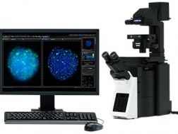
Providing intuitive operations and a seamless workflow, cellSens software’s user interface is customizable so you control the layout. Offered in a range of packages, cellSens software provides a variety of features optimized for your specific imaging needs. Its Graphic Experiment Manager and Well Navigator features facilitate 5D image acquisition. Achieve improved resolution through TruSight™ deconvolution and share your images using Conference Mode.
- Improve experiment efficiency with TruAI™ deep-learning segmentation analysis, providing label-free nuclei detection and cell counting
- Modular imaging software platform
- Intuitive application-driven user interface
- Broad feature set, ranging from simple snapshot to advanced multidimensional real-time experiments
