FV4000
 | Transforming Precision ImagingEmpower Your Confocal Microscopy Imaging ExperimentsTransform your images with the new FLUOVIEW™ FV4000 confocal laser scanning microscope. Advanced imaging technology enables higher precision images to empower researchers with more reliable data from their samples. With our breakthrough SilVIR™ detector at the core of the system, achieve much lower noise, higher sensitivity, and improved photon resolving capabilities. With the FV4000 confocal microscope, researchers can acquire higher-quality, quantitative image data in less time and with less effort. |
|---|
Experience the systems innovations, including:
*As of October 2023. |
Easy-to-Acquire, Quantitative Confocal DataThe FV4000 confocal microscope uses our advanced, silicon-based SilVIR™ detector that makes it easier than ever to acquire precise, reproducible data. SilVIR Next-Generation Detector Technology The SilVIR detector combines two advanced technologies—a silicon photomultiplier (SiPM) and our patented* fast signal processing design.
*Patent number US11237047 Learn more about the SilVIR detector |
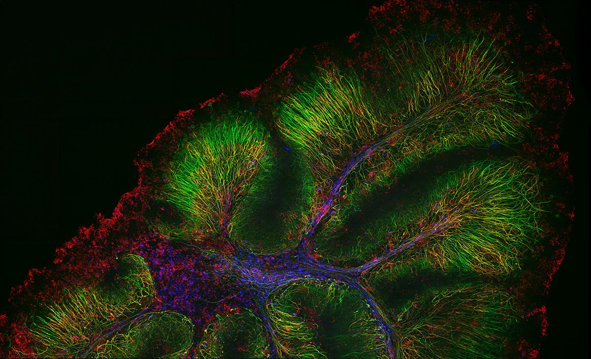
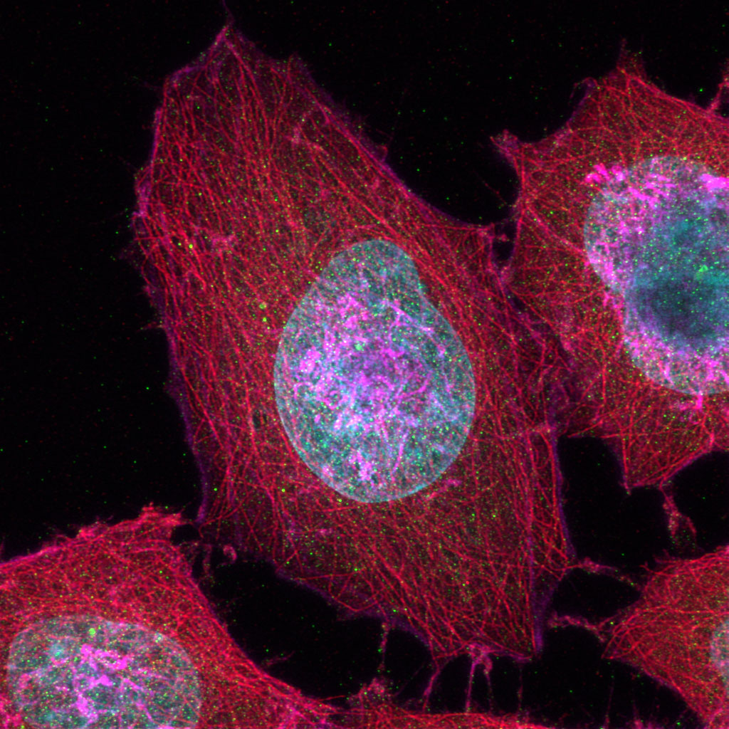
Neurofilament-heavy chain (NFH) in green, myelin basic protein (MBP) in red, glutathione S-transferase pi 1 (GSTpi) in blue. Mouse cerebellum captured with a UPLXAPO40X objective.
|
|
|
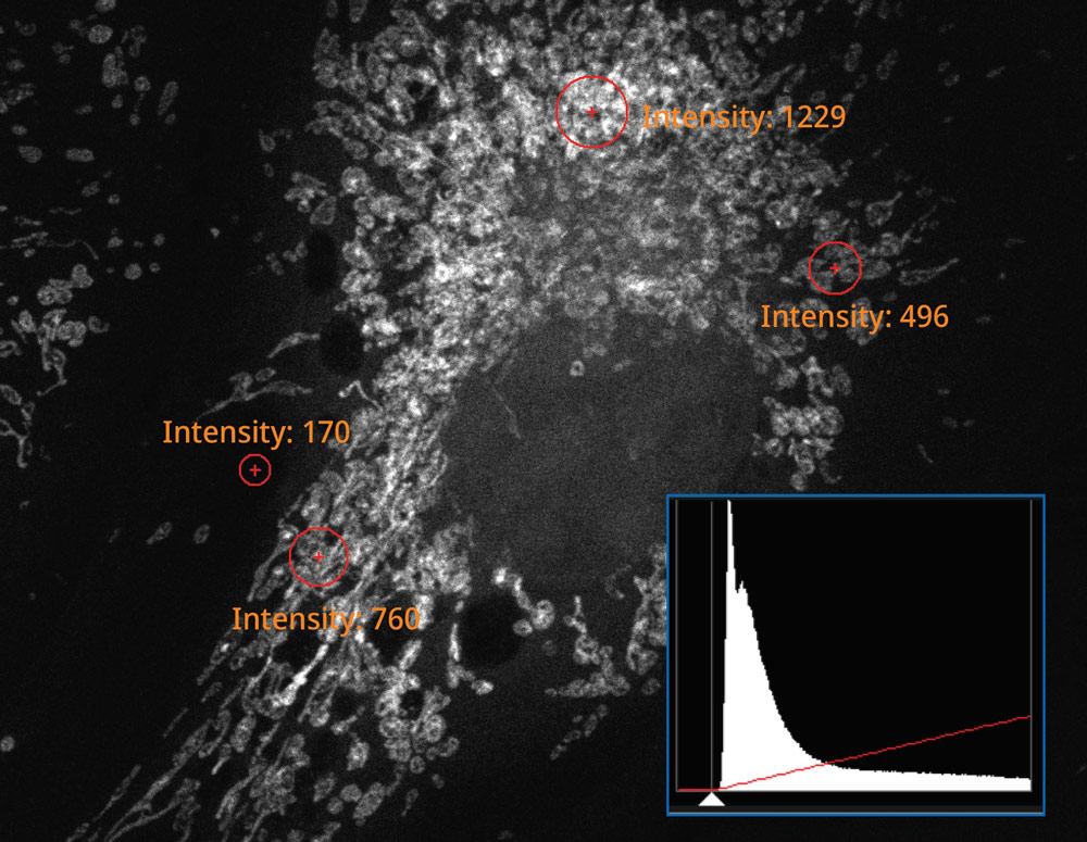
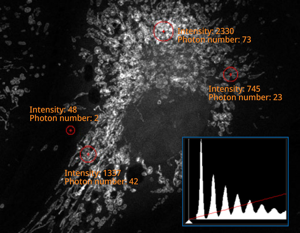
The histogram on the image captured using the SilVIR detector shows a discrete pattern where the intensity can be converted to the photon number. The detector’s fluorescence intensity can be quantified as the photon number, and the background level is extremely low. |
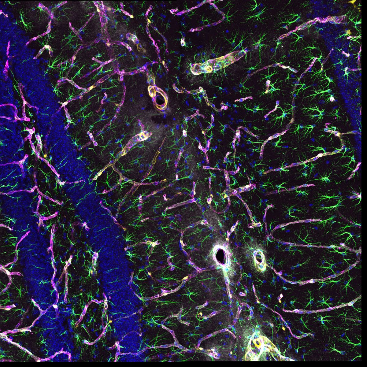
More Information from Your Confocal ImagesThe system’s updated TruSpectral technology combined with high sensitivity SilVIR detectors enable you to see more by making it possible to multiplex up to six channels simultaneously.
|
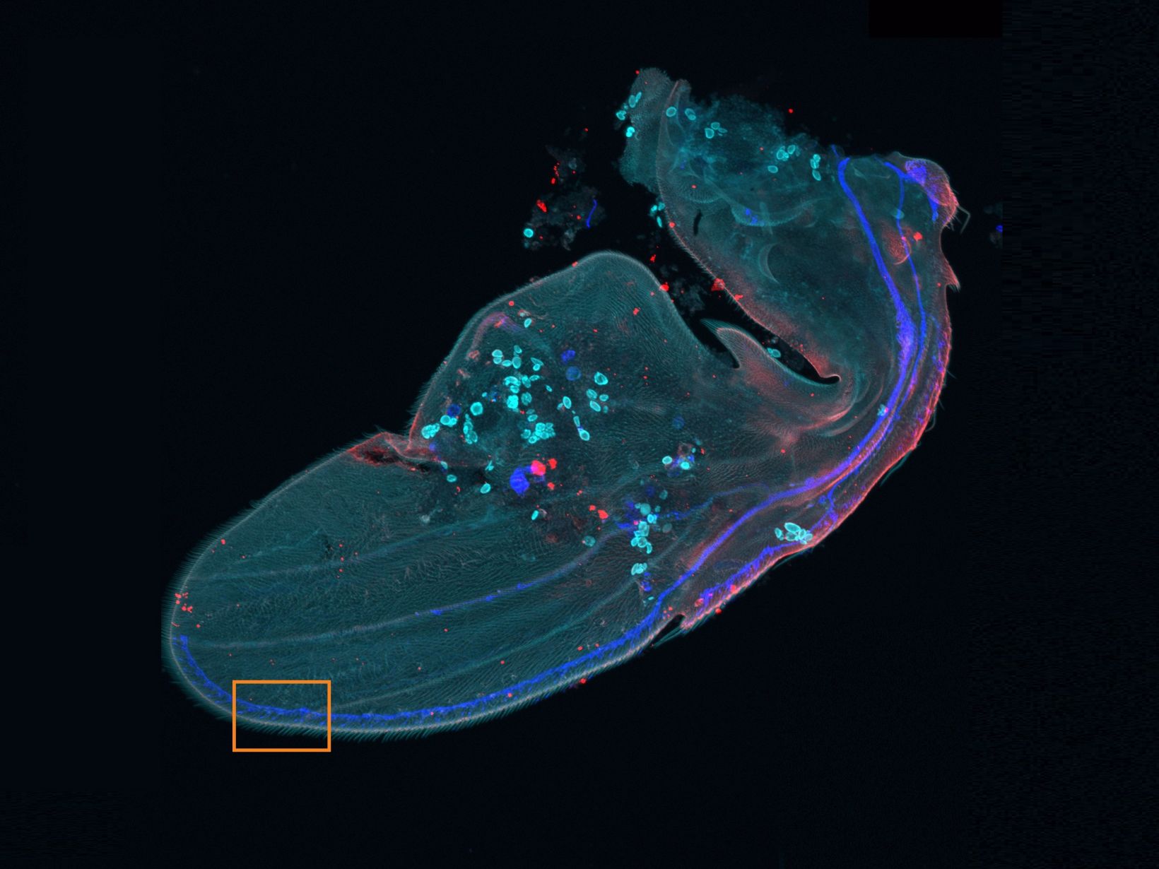
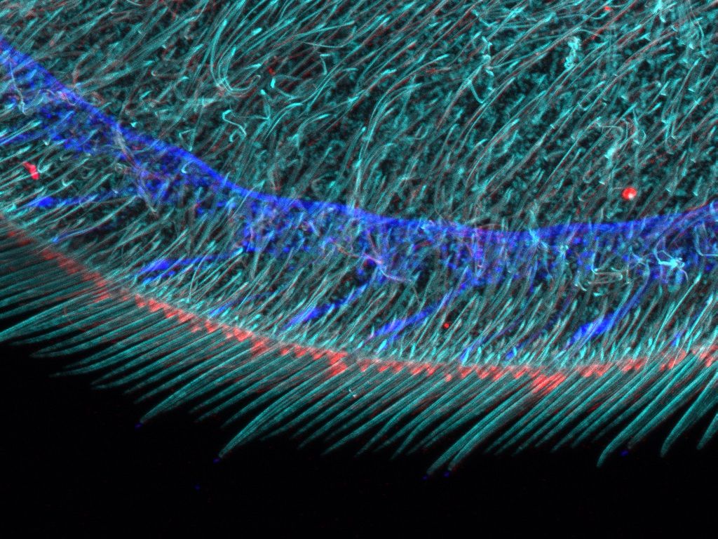
Flexible Macro to Micro ImagingThe macro-to-micro workflow enables you to easily observe the target sample from the macro level—whole body or tissue—down to the cell or subcellular level.
| ||||
Gentler High-Speed Time-Lapse Confocal Imaging | |
Related VideosHeLa cells labeled by MitoView 720. XYZT imaging by 1K resonant scanner for 30 min. | Time-lapse imaging is easier with smart features:
|
Reproducible Image Data Between Users and SystemsThe SilVIR detector has less sensitivity loss over time compared to previous-generation detector technologies. With our laser power monitor (LPM) and TruFocus™ Z-drift compensator, achieve reproducible images under consistent conditions for better reproducibility. Different users on different days can acquire the same precise images using the same settings. Even the images acquired by different FV4000 microscopes can be compared and discussed using the same photon number intensity scale. |
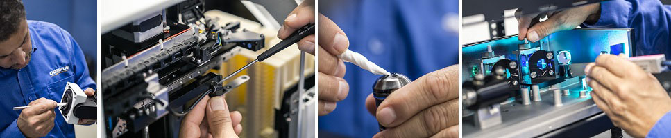
Microscope Support and Service You Can Count OnWe designed the FV4000 system to be easy to maintain:
We stand behind our products with a commitment to fast service and technical support. We offer various support plans to keep your microscope running at peak performance at a predictable cost as well as remote support options, so you don’t need to wait for an engineer or specialist to visit if you’re having an issue.
|
Need assistance? |
Sorry, this page is not
available in your country.