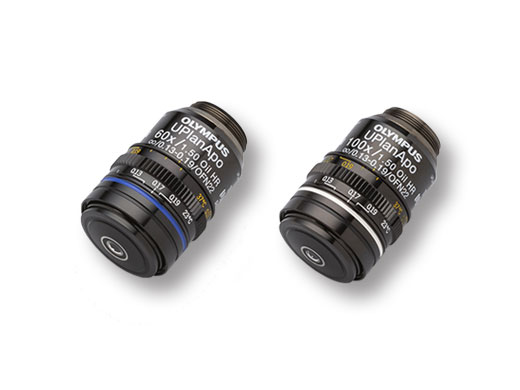
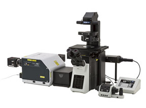
High-Resolution Objectives for Super ResolutionA high numerical aperture (NA) is important for super resolution images.Our proprietary polishing technology enabled us to create the world’s first plan-corrected apochromat objectives with an NA of 1.5*1. Combining this objective with the IXplore SpinSR system improves the brightness and resolution of your super resolution images. The objectives are particularly useful when visualizing surface microstructures. *1 As of Nov. 2018, According to Olympus research. |  |
Scale bar: 200 nm | Super ResolutionResolve confocal images using the confocal technique and Olympus super resolution (OSR). Green: Alexa488 labeled Nup358 which localizes to the cytoplasmic surface of nuclear pore complexRed: Alexa555 labeled Nup62 which localizes to the nuclear pore complex central plug Localization of Nup358 and Nup62 can be distinguished by super resolution technique. *Nuclear pore complex of HeLa cell |
|---|
High-Resolution Objectives Selection Guide
|
Working Distance
(mm) | Magnification | Objective Field Number*2 | Numerical Aperture | Immersion | Applications | |
|---|---|---|---|---|---|---|
| UPLAPO60XOHR | 0.11 | 60X | 22 | 1.50 | Oil | Real-time, super resolution imaging for live cells/super resolution imaging of tiny structures, such as organelles/whole cell TIRF imaging |
| UPLAPO100XOHR | 0.12 | 100X | 22 | 1.50 | Oil | Real-time, super resolution, imaging for live cells/super resolution imaging of tiny structures, such as organelles/high-resolution imaging of cell membranes or subcellular organelles, and single-molecule level experiments |
*2 Maximum field number observable through eyepiece
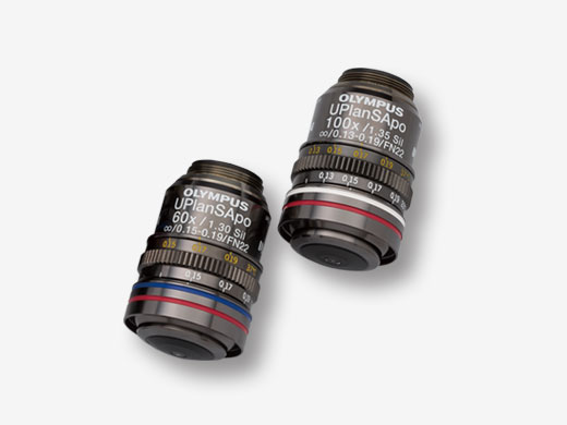
Silicone Immersion ObjectivesSilicone immersion objectives are optimized for live cell and live tissue imaging. By properly matching the refractive index, images are clearer and brighter, and time−lapse observations become more reliable and less complex because silicone oil does not dry at 37 °C (98.6 °F). With a high numerical aperture and long working distance, these objectives, when combined with Olympus super resolution, enable you to observe microstructures on your sample's surface as well as deep inside it. For example, both molecule localization and nerve cell microstructures can be observed with high resolution. |  |
Related Videos | 3D Time-Lapse of a NeuronObtain detailed three-dimensional super resolution image data during time-lapse imaging. |
|---|
Silicone Immersion Objectives Selection Guide
|
Working Distance
(mm) | Magnification | Objective Field Number*3 | Numerical Aperture | Immersion | Applications | |
|---|---|---|---|---|---|---|
| UPLSAPO100XS | 0.2 | 100X | 22 | 1.35 | Silicone oil | High-resolution for subcellular imaging |
| UPLSAPO60XS2 | 0.3 | 60X | 22 | 1.30 | Silicone oil | High-resolution and long-term, time-lapse imaging of single cells |
*3 Maximum field number observable through eyepiece.
Related Products
IXplore SpinSR |
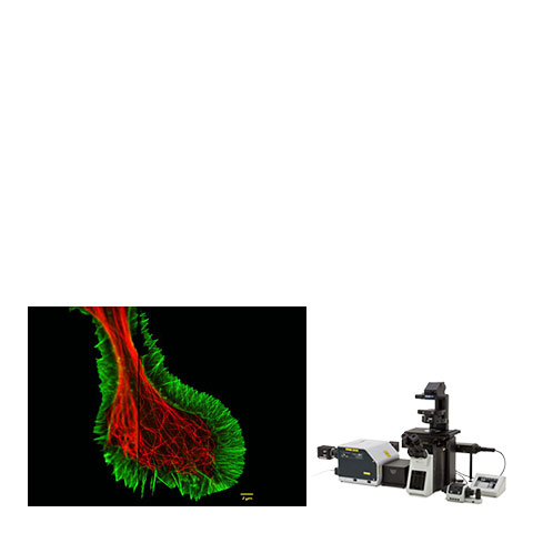
*Banner Image: Fluorescent staining of microtubules (red: Alexa 594) and actin (green: Alexa 488 phalloidin) in growth cone of NG108 cells.
By courtesy of: Dr.Kaoru Katoh , Biomedical Research Institute,National Institute of Advanced Industrial Sciences and Technology
Sorry, this page is not
available in your country.
Sorry, this page is not
available in your country.


