We love to showcase the amazing work being done by Olympus customers over at the Olympus Life Science Instagram account.
This past month featured our first-ever Instagram Takeover, with Håkan Kvarnström sharing a week’s worth of images and answering questions about his work.
The winner of Olympus’ 2018 European Image of the Year Europe Award, Håkan is experienced capturing the beautiful side of science. As he describes, his work is “about the images and the stories they tell. The micro world is filled with exciting and beautiful discoveries.”
It’s no surprise that the top five most popular images this month all came from his takeover. Check out our February top five below with Håkan’s captions.
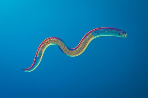
This photograph shows a nematode discovered in a sample collected from swamps in Florida. It is estimated that up to 80% of all individual animals found on earth are nematodes, making them the most abundant animal on earth. They occupy almost every ecosystem, including marine water, freshwater, and soils from polar regions to the tropics.
Just like general photography, lighting techniques are vital in making striking images. I believe beautiful images have a significant impact on conveying the stories they carry. Therefore, it is crucial to master different lighting and contrast techniques to best illuminate the subjects under the microscope. Many microscopic life forms are transparent and have no color, making them nearly invisible if illuminated with a plain light source. To increase contrast and make transparent subjects visible, various contrast-enhancing techniques are used. These techniques modify light in different ways, creating pseudo- colors, shadows, colored or black backgrounds, or even 3D-like textures. Contrast-enhancing techniques can be very expensive, but the good news is that one of the most useful methods in photomicrography is very inexpensive. You can create colorful images by adding a simple polarized filter below the condenser and a second filter in the pathway right after the light has passed through the objective. Differences in thickness and refractive indices will produce different colors, while turning and moving the specimen (or filters) will create different effects. While some microscopes are factory equipped with polarized filters, almost any microscope can be equipped with a polarized filter by cutting the plastic lenses from a pair of 3D-movie glasses to fit the microscope.
Image captured using an Olympus BX51 with a UPLSAPO40X objective using polarized light.
Although my work began as an interest in microscopes, it has become more about the images and the stories they tell. The micro world is filled with exciting and beautiful discoveries. I put considerable effort into portraying these life forms as aesthetically as I can. I view myself as a nature photographer, documenting life invisible to the naked eye. When you look around, you see organisms visible to the naked eye—trees, plants, birds, and other wildlife. For a long time, scientists thought that nature was made up of only these visible things. We have since learned that two-thirds of life on earth are microorganisms that cannot be seen without a microscope. Modern microscopes with digital cameras have revolutionized scientific photography and made these worlds accessible.
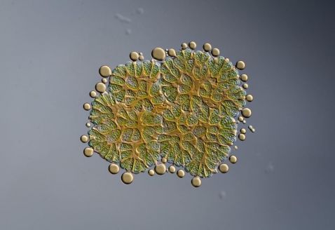
Today’s photograph shows a green alga called Botryococcus braunii. 30–40% of the cell’s biomass consists of oil, which can be seen leaking out around the edges of the cells. It was captured using an Olympus BX51 microscope with a UPLSAPO60XW water immersion objective and differential interference contrast (DIC). A great advantage of the water immersion objective is that its working distance is much higher than the corresponding oil immersion objectives. This is an advantage for thicker samples in wet mounts where the subjects can move in the water sample. This subject was larger than the field of view of the objective, so I had to stitch the final image together from images from the top part and the bottom part, respectively.
Image captured using an Olympus BX51 microscope with a UPLSAPO60XW water immersion objective and differential interference contrast (DIC).
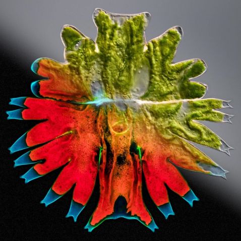
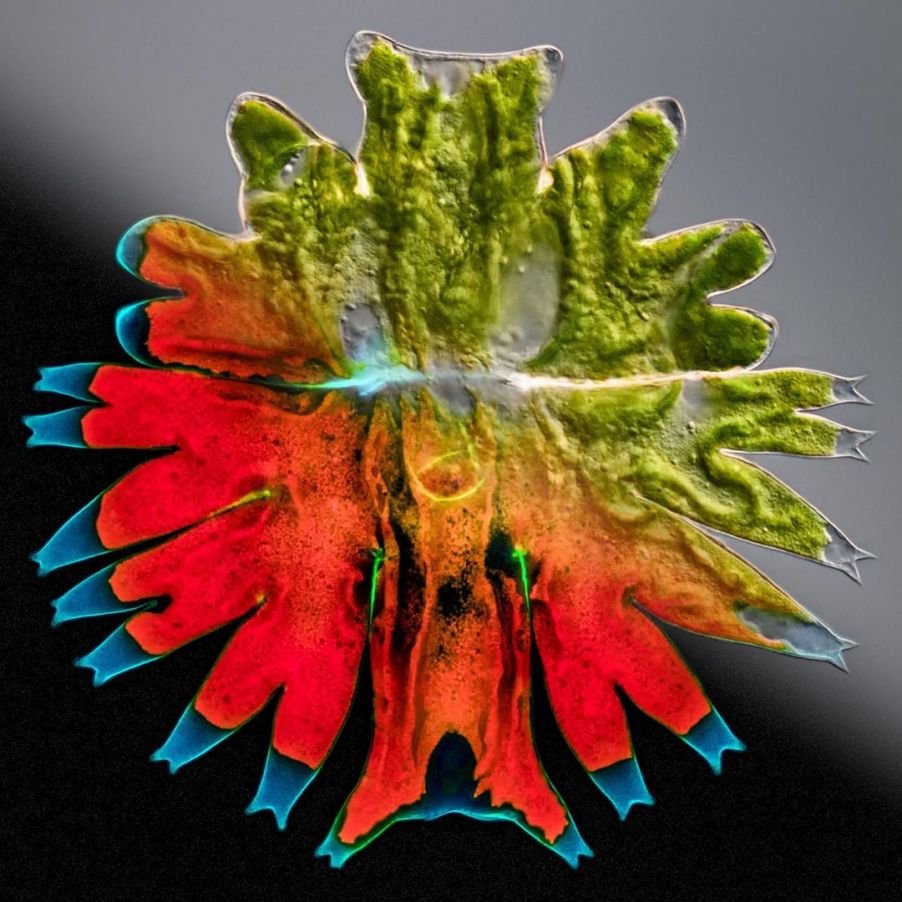
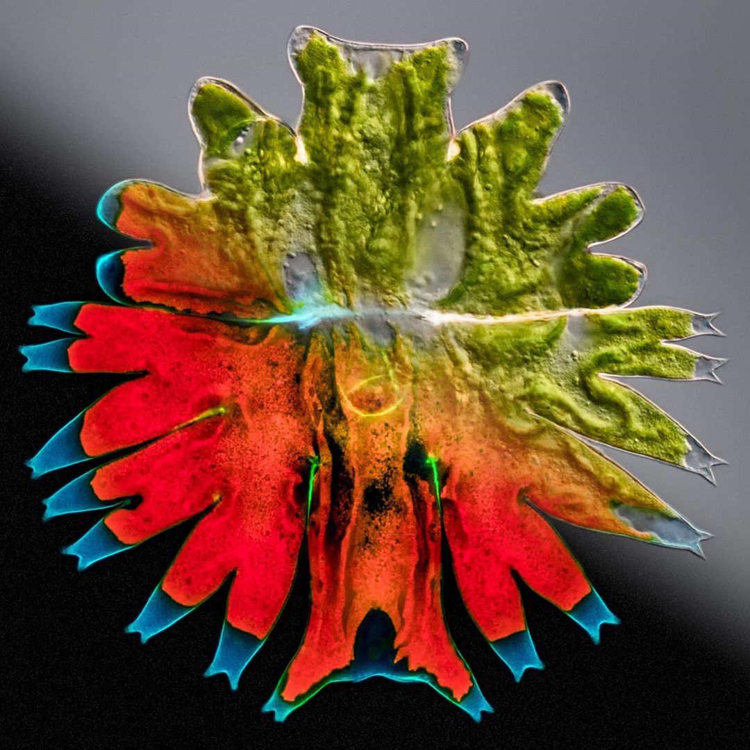
This image was inspired by the well-known photographer, Stephen Wilkes (@stephenwilkes) and his series of images called “Day to Night” where both day and night were captured in a single image.
Science photographers can learn a lot from traditional photographers, e.g., image composition, proper use of light, and artistic expression. What is fascinating with microphotography is that different lighting and contrast techniques highlight different features of the subject under the microscope. Some highlight the surface and give a good idea of the exterior shape of the subject, whereas other techniques cut like a knife through the subject to visualize interior structures.
Some images in this series are made of up to 100 images stacked, stitched, and then combined in photoshop to achieve a blended image combining the two illumination techniques. The challenge with this technique is to work fast enough behind the microscope because any movement of the subjects will ruin the final image.
Image of a green alga, Micrasterias, captured using DIC and fluorescent illumination to produce a composite of two images.

This photograph shows two different species—a diatom, Tabellaria fenestrata var. asterionelloides, and a desmid, most likely Staurastrum pseudosebaldi. The Staurastrum has the classical green color from its chloroplasts. Diatoms still have chloroplasts but also contain other pigments such as fucoxanthin, giving them a brownish-yellow color.
I often get questions about how I make my images and the workflow I use. Digital cameras have revolutionized photomicrography, in turn, creating many techniques to improve an image that used to be virtually impossible using analog film. I’m going to discuss the most important technique I use—focus stacking. Focus stacking is a technique to overcome the problem with the extremely shallow depth-of-field of a microscope objective. Using high NA objectives at high magnification, the depth-of-field may be fractions of a millimeter. That means that if the subject has a thickness of a micrometer, only a small part of the subject will be in focus at the same time. Moving the specimen or objective up or down is necessary to shift the areas in focus. A typical stack may be 10–20 images in which different parts of the subject are in focus. In some cases, a stack may consist of up to 100+ images where only fractions of the pixels are used from each image to form the final image. The focus stacking software is not only about the selection of pixels in focus. Equally important is the selection of pixels not in focus, especially if you want a clean background. A great technique is to take a few images of a highly out-of-focus subject just to capture a clean background. Then, during stacking, the software is used to select the pixels you want for the subject (in focus) and the background (out-of-focus). The stacking software helps you select the right pixels through intelligent algorithms.
The image is a composite stack of 43 images. Captured using an Olympus BX51 with a UPLSAPO60XW water immersion objective and DIC.

The final image shows a small crustacean, Simocephalus vetulus, another specimen inspired by Stephen Wilkes (@stephenwilkes).
Image captured using brightfield and fluorescent illumination to produce a composite of two images.
A big thanks to Håkan Kvarnström for sharing so many wonderful images with us and providing such wonderful details about his microscopy techniques.
To see more images like these, be sure to follow us on Instagram at @olympuslifescience!
Interested in sharing your own images?
Visit our image submission site.
