Allowing the unseen to take flight
In Marko Pende’s experience, most people are afraid of insects to a certain extent—except for ladybugs, which most people see as harmless and friendly. We have all seen ladybugs on a summer day with their familiar round red shape. What most of us never see, however, is a ladybug at large scale and with its wings open.
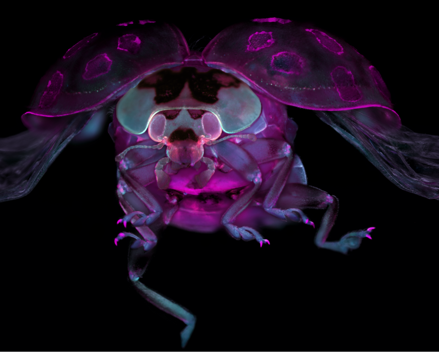
This is what Pende found most interesting about using a ladybug as a model for microscopic imaging—the idea that he could create an image that would challenge the way people see and think of a common American insect, and give them a new perspective on the complex physical makeup of a creature that appears very simple to the naked eye.
To create his winning image, Pende worked hard to position his sample in such a way that it appeared in flight, furthering the emotional impact of his image, as most of us do not typically get the chance to view a ladybug straight in the eyes as it flies toward us.
Pende was selected as Evident’s 5th Annual Image of the Year Award Americas winner for his fascinating image of a tissue-cleared ladybug. He was awarded his choice of an Olympus CX23 upright microscope or SZ61 stereo microscope for his winning submission.
Congratulations on being named our Americas winner! What does your winning image show us?
This image shows us a tissue-cleared ladybug stained with DAPI and TO-Pro dyes and injected with 70kD 555 Dextran.

Evident Image of the Year Americas winner: Tissue-cleared ladybug. Captured by Marko Pende.
Why did you choose this image as your entry for the competition?
Many people are afraid of insects, but when they see a ladybug, they are usually not afraid of it. For me, this was a nice image, something positive that I wanted to record. And I also felt that most people have probably never seen a ladybug in this particular way. So I found it very interesting to use as a model.
What did you find personally exciting about this image?
In general, I am interested in all kinds of imaging—photography and interesting motives behind image choices. The ladybug was a very intriguing choice for me. I wanted to show a ladybug in a way that was never seen before.
How did you create this image?
I used a stereo microscope with 22x magnification. The imaging technique is the interesting part—this was not a transparent sample, and what I do in my career is develop tissue-clearing methods, which means I make samples transparent. What I wanted here was to make the ladybug semi-translucent—if you were to make it completely transparent, you would lose a lot of the interesting shapes in its structure. So what I did was label it with nuclear dyes. I used TO-Pro and DAPI dyes in different colors to create some differences in the staining pattern, and I injected the ladybug with Dextran to achieve a bit of a different body color.
The positioning of the ladybug was tricky because I was using a top-down objective. So what I did was take a jar and filled it with water and put the ladybug on a needle so it was facing upwards toward the objective. I wanted to generate the feeling of the ladybug flying even though it was static.
How did you discover the sample you used to create this image?
I was not allowed to sacrifice a ladybug because my girlfriend would have been so mad at me! So I had to wait to find a dead ladybug that was recently deceased and had not begun to dry out and disintegrate.
Does this image help benefit scientific research?
I think it helps to raise general public interest in science, and it helps to associate science with a positive topic. I believe that is a general benefit.
Marko Pende began looking through the lens of the microscope at age 10. Today, he specializes in both underwater photography and microscopic imaging.
When did you first learn to use a microscope?
My grandfather was a professor of immunology and biochemistry in Croatia, and he did microscopy there. At the age of 10, I got my first microscope from him. It had no internal lights, so you had to put a light bulb in it. From a very young age I started microscopy.
How long have you been creating art with a microscope?
In 2018 I published a paper in Nature Communications with some of my images and then was invited by the Genetics Society of America to participate in an imaging contest. I didn’t win, but I did get an honorable mention. So that was the first time I started to think, maybe I can participate in artistic imaging contests. So I would say that for 7 or 8 years I have been creating images purely for art’s purpose.

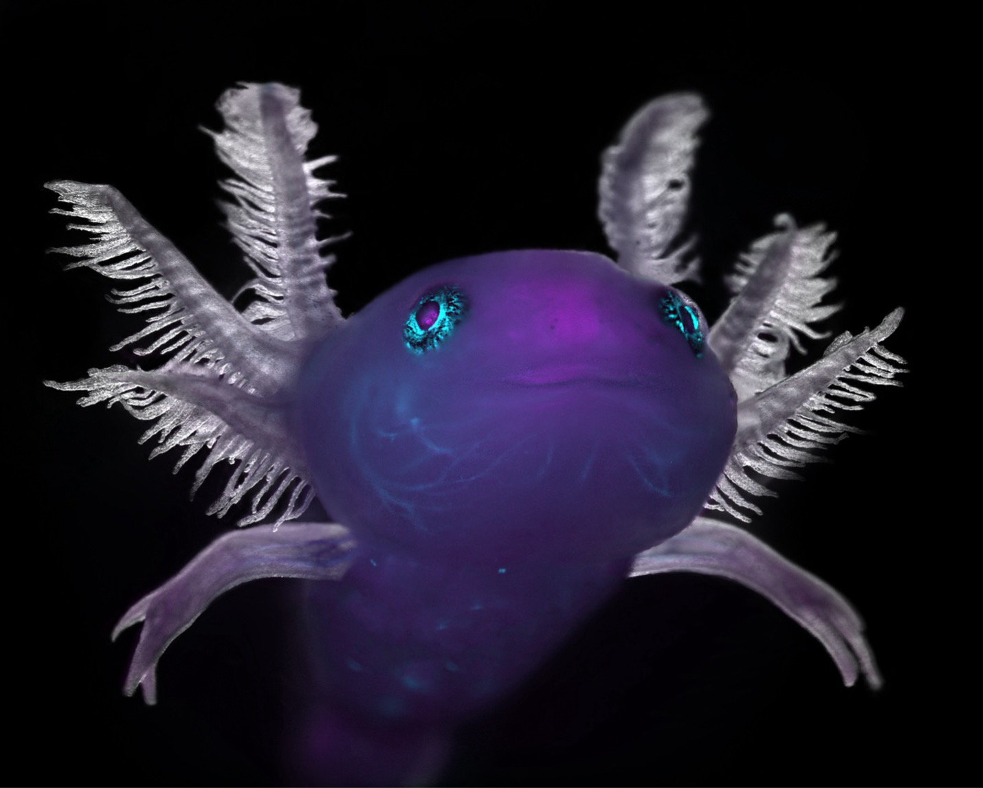
Axolotl captured with a stereoscope. Captured by Marko Pende.
What do you find most fascinating about microscopy?
Making the invisible visible and showing people what they usually wouldn’t see. I use a microscope like a photo camera, especially stereoscopes, and I am very thankful for this.
Where do you think this fascination stems from?
I was very encouraged by my family in general to explore art as a child. We had a yellow analog underwater camera that I would use as a child, and I was always interested in photography. It’s something I have always enjoyed a lot, and a microscope is just a different way to create images, of course on a much smaller scale. I have done underwater photography for many years, so I like to create images, especially microphotography. I like to make pictures of what I call weird little creatures.

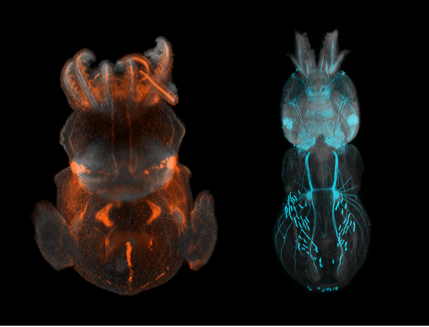
Squid creatures captured with an Olympus 4X objective and a custom light sheet. Captured by Marko Pende.
What do you do professionally? Does your profession intersect with imaging?
Currently I am a postdoc, and my primary work is in the development of tissue clearing methods, which includes work with amphibian embryos and mice. I earned my PhD in biomedical engineering from the Vienna University of Technology. I have worked with custom microscopes for 11 years. My whole career is based on imaging and developing methods for imaging, but it’s also my hobby, something I really enjoy.
What kind of experience do you have with Evident and Olympus microscopes?
I have extensive experience with Olympus. My entire PhD was imaged with Olympus objectives. We had a custom-built light sheet system, with Olympus objectives optically corrected for immersion. My favorite objective was the XLFLUOR4X, with an NA of 0.28, that we custom-corrected for immersion into solutions. This is an excellent objective. I also currently use an SZX16 stereo microscope quite a bit, which is a workhorse of a microscope for fast checking of fluorescent signals. I have had a very good experience with Olympus in general.
If you could image anything next, what would it be?
My sister would kill me for saying this, but a juvenile octopus would be really cool! Sometimes I also just have an idea that pops up and I visualize in my head what it should look like. And then I try to find the sample that matches my vision. I also get inspired by others. I also think that creating an image of the face of a tardigrade would be super cool, or maybe some crazy shrimps. If I could ever image a hairy frog fish, that would be very cool—this is more the diver in me speaking!


