The beauty of individual forms becomes a new singular experience
While looking through a batch of old sample slides during the early days of COVID lockdown, Daniel Han discovered some fossil diatoms that captured his interest. After experimenting with different ways of imaging them, Han said he became “fascinated and obsessed” with their inherent beauty and the qualities that make them a truly unique subject to work with.
In working with diatoms—and adding more diatom samples to his collection—Han unlocked a new sense of curiosity around their endless variety of shape, form, and texture. As he discovered new ways to create colorful, dramatic diatom images under the microscope, he also became inspired to start arranging multiple diatoms into shapes of their own, creating one-of-a-kind expressions made up of microscopic art within art.
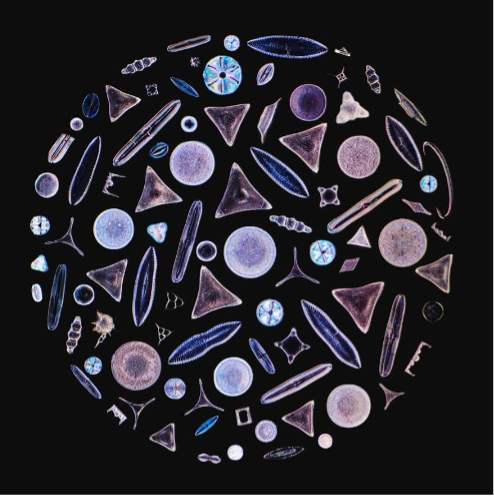
Han, of Australia, was selected as Evident’s 5th Annual Image of the Year Award Asia-Pacific winner for his dramatic image of arranged micromanipulated diatom frustules.
In recognition of his winning submission, Han received his choice of an Olympus CX23 upright microscope or SZ61 stereo microscope.
Congratulations on being named our Asia-Pacific winner! What does your winning image show us?
This image showcases arranged micromanipulated (mostly) diatom frustules from a mixture of localities.
Evident Image of the Year Asia-Pacific winner: Arranged micromanipulated diatom frustules. Captured by Daniel Han of Australia.
What did you find personally exciting about this image?
Diatoms come in many different shapes and forms. I call them nature’s glass gems. Among the arrangement were also a few silicoflagellates.
How did you create this image?
To create this image, I used a custom darkfield setup at 20X (10X with x2 magnification changer) and stitched two stacked images together to complete the circle.
How did you discover the samples you used to create this image?
I initially discovered diatoms during the times of early COVID among a batch of old slides I had picked up. I have been fascinated and obsessed with them ever since. Fossil diatoms from different localities come in different shapes and sizes, and they are truly a unique subject.
Did you face any challenges when creating this image?
There were in fact a few challenges.
When it came to depth of field, diatoms are thick, but the plane of focus was thin. A single image will only showcase a thin layer of features. Z-stacking was used to overcome this.
In regard to contrast, diatoms are glass frustules; they have a similar refractive index to the coverslip and the slide, thus inherently lacking contrast. To increase contrast, high refractive index mountant was used (~1.75RI), and I chose to utilize the technique of darkfield microscopy combined with polarized light. The colors seen in the image were from interference, as the frustules themselves were opaque.
The condenser I used lacks darkfield capability, so I designed a tray that slides above the polarizer that holds a darkfield patch stop.
I needed to achieve adequate FOV. My 10X (20X with x2 magnification changer) only captured half of the arrangement. To fix this, I stitched two Z-stacks together.
Why did you choose this image as your entry for the competition?
When entering a competition like this, I usually choose a few images using different techniques and I always make sure that at least one of the subjects is made up of diatoms. I chose this specific image because it was rich in forms and I was satisfied with the performance of my custom-designed parts.
Is there a message inspired by this image?
Diatom imaging is an art form, and many people think that expensive imaging modalities such as differential interference contrast are required to successfully create striking diatom images. I think darkfield and brightfield imaging are just as valuable and produce beautiful results.
Does this image help benefit scientific research? If so, can you explain how?
There is a lot of research pertaining to diatoms, fossil diatom deposits, and different localities. My arrangement used freshwater fossil frustules from various locations. For aesthetic purposes, a few sponge needles were added in as well. These were found among the same samples.
Diatoms are examined in a variety of ways, including as a means to assess water quality changes to determine the effectiveness or assess the requirements of conservation efforts.
When did you first learn to use a microscope?
I first learned how to use a microscope during COVID-19 lockdowns when I picked up a decommissioned Olympus BX40 for a cheap price and fixed it up. I come from an electrical engineering background; however, I did construct a metallurgy microscope for my thesis project that was focused on the automation aspect of image acquisition. I also did one course in optics and worked in a photonics laboratory as a researcher for a while. This gave me a baseline understanding of the technical aspects of microscopy.
What first gave you the idea that microscopes could be used to create art?
When I acquired my first microscope during COVID, I also had online classes. I chose to leave some of my easier subjects until the last year of my studies but this decision backfired. Being in lockdown, it was often very boring. I started using my metallurgy setup to take photos of subjects such as butterfly/moth wings and other oddities, but a magnification of 5X-10X could only go so far. After I picked up a batch of vintage microscope slides online, I discovered a wide range of fascinating microscopy subjects. That was when I began to create microscopy art. I might not know a lot about the subjects, but I can certainly showcase their beauty!
How long have you been creating art with a microscope?
I would say around five years for sure. The metallurgy setups I used in the past certainly count as homebrew microscopes, so maybe even around 10 years. I started this hobby in my late high school years.
What do you find most fascinating about microscopy?
I like to try out different imaging techniques on the same subject to see the different results. Certain techniques are often not officially offered, or are very costly. This prompts me to design parts to achieve the results I want. The winning image is a good example of that.
Where do you think this fascination stems from?
It is mainly driven by curiosity and challenging myself. I feel an overwhelming sense of satisfaction if I can design and produce something that matches, if not surpasses, official offerings for the specific purpose of microscopy art. The components offered are usually targeted at the needs of scientists and industrial users rather than those who intend to create art.
What do you do professionally?
Professionally, I work as a project manager and supervisor.
Does this profession intersect with imaging? Or is this more of a hobby, art form, or passion for you?
This is more of a hobby for me. I do occasionally visit sites as part of my job, and if I know that there are bodies of water or other notable microscopic interests onsite, I will often bring a field kit to acquire some samples while I am there. While water life films are not my specialty, I do like recording videos and sharing them.
What are you currently working on from a microscopy standpoint?
There are multiple concurrent projects I am working on for microscopy. Mainly, I have been disappointed by the transmitted light illuminator (halogen, a bit dim and requires frosted glass) and LED alternatives I have tried. Hence, I will be designing an LED-based solution that is bright enough to replace the halogen lamp while being able to achieve Köhler illumination. I do want it to be fanless as fans induce visible vibrations. This is somewhat challenging because most solutions I have tried fail to surpass 100W halogen lamps in terms of brightness.
What kind of experience do you have with Evident and Olympus microscopes?
I have worked with Olympus/Evident during a past job and purchased lots of items for my microscopes as well. Evident has always been professional and responsive to inquiries. Any issues were sorted very quickly.


