Spheres, snow, and silverfish. Alliteration, and the selection of images that made the final monthly top 5 for the year!

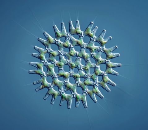
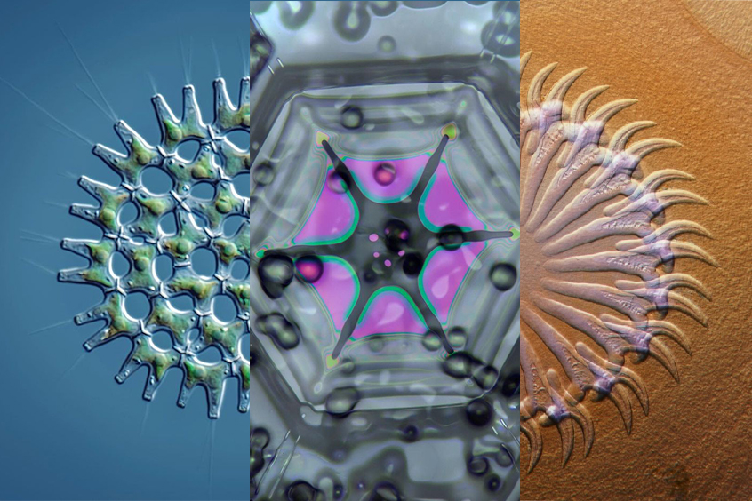
No, this isn't a green coronavirus. This round organism is actually a tiny green algae of the genus Pediastrum, collected in Vombsjön, Sweden. What's interesting in this image are the flagella visible at the end of each “arm.”
Description and image courtesy of Håkan Kvarnström.

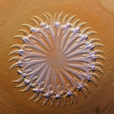
Another striking circle captured enough people’s attention to make the Top 5 this month. This image shows the head of a tapeworm (Taenia pisiformis) from a brown hare.
2019 Image of the Year Submission. Captured on an Olympus BX51 microscope. Image courtesy of Dr. David Maitland.
Submit your best 3 images by January 31, 2021 for a chance to earn the 2020 Image of the Year title and win a SZX7 stereo microscope with a DP27 digital camera!

In the northern hemisphere, winter has arrived, and with it (in many parts of the world) comes snow. In December, we were thrilled to feature our latest Instagram takeover, celebrating the first day of winter. Over three days, Linden Gledhill shared his images and techniques for imaging snowflakes.
“I've been fascinated by these stunningly beautiful creations since I first set eyes on them as a child. Their forms have captured the hearts of people and are so symbolic of the winter season.
I often get asked, “How do you plan snow crystal shoots?” For starters, I pay close attention to the weather forecast. Significant snowstorms aren't really that common where I live just outside Philadelphia. Most of the time I work at night when the temperatures are the lowest as it has to be at least -5 °C (23 °F) to have any chance of success. Next, I have to decide which microscopy technique and Olympus systems I should use—a transmitted light Olympus BH2-BHS fitted with S-plan achromats and DIC lighting or a darkfield/reflected light Olympus BH2-BHT fitted with NeoSplans and a BH2-UMA illuminator. Both microscopes have been modified to use LED lighting, high-speed flash, and focus stacking. I place the chosen equipment on a bench in the mouth of my garage and open the windows to ensure that the room cools down to the external temperature. I then bundle up in warm clothes and wait for the snow to arrive!
Today’s image gallery was captured using the darkfield anulus in the NeoSplan objectives and the BH2-UMA illuminator with high-speed flash. The very shallow angle of light brings the very edges of the crystals and makes them look translucent. Refraction can also be seen, and small air cavities are revealed within the main body of the crystals. The overall visual impact is glass like.”
Captured in darkfield on an Olympus BH2 microscope. Images courtesy of Linden Gledhill.

It’s no surprise that Linden Gledhill’s stunning snowflakes and explanations placed him in two of the top 5 spots for the month!
“Today I will explain how snow crystals are collected and transferred to the microscope!
Finding a perfect crystal is like panning for gold; it takes many hours and considerable determination. I use a black foam board to capture the falling snow. Transferring snow crystals to the microscope can be tricky. I use a statically charged plastic toothpick to transfer single crystals. If I'm doing transmitted light methods, the crystal goes onto a slide. If I'm imaging with reflected light, I place the crystal on a 1 cm platform with a felt surface. The platform is supported by a stem with a ball joint, which allows me to level the crystal so it's exactly at 90 degrees to the epi illumination. My BH2-UMA illuminator is modified with an LED light source and a flash system being directed by a beam splitter. This helps gain alignment with very low LED lighting and then switch to flash for the final imaging.
Imaging has to be done very quickly due to sublimation, a process where the ice converts to water vapor directly without melting. As soon as you capture the crystal and place it on the microscope, you have changed the ambient vapor pressure, and sublimation starts to occur. If the crystal is too big for the field of view, I'll create a multi-frame panorama. I have been known to work all night if the storm is producing nice crystals. This often results in different crystal forms as the atmospheric conditions change!
The natural interference images you see here are captured using NeoSplan objectives and the BH2-UMA illuminator with high-speed flash and direct epi-lighting. People are often surprised by the extremely bright colors that can be seen using this method. This is due to light bouncing back from multiple surfaces within the crystals, causing constructive and destructive interference. This happens when the faces are separated by gaps, which approach the wavelengths of visible light. These little gems are very rare, but when I find them, I get goosebumps because they are unbelievably beautiful. Snowstorms showing these forms will keep me hunting for more all night!”
Captured on an Olympus BH2 microscope. Images and caption courtesy of Linden Gledhill.

Fun fact: Silverfish (lepisma saccharina) can outrun most of their predators when on horizontal surfaces but lack agility and speed when climbing vertical surfaces. Thankfully, this makes them easier to catch when found in a bathroom sink!
2019 Image of the Year Submission. Captured on an Olympus BH2 microscope. Image courtesy of Marco Jongsma.
As we leave the year behind, we’re thrilled to share the overall top image of 2020:

This little crab spider may not be smiling, but it sure made us smile! Many members of the Thomisadae family are also known as flower spiders, which makes it no surprise that this little fellow was collected from a flower garden in the Netherlands.
Captured on an Olympus BH2 microscope at 10x magnification. Image courtesy of Marco Jongsma.
To see more images like these, be sure to follow us on Instagram at @olympuslifescience!
Interested in sharing your own images? Visit our image submission site.
Submissions are now also being accepted for our 2020 Global Image of the Year Award Competition. Click here to learn more and see and how you can win a SZX7 stereo microscope with a DP27 digital camera or CX23 microscope.
Related Content
Diatoms to Daphnia: Our Most Popular Microscope Images for November 2020
5 Ways to Extend the Life (and Performance) of Your Microscope
The Art and Science of Neurogarden: Meet the 2019 IOTY Global Winner


