The end of the year always sneaks up on us, and with it the holiday season. Our top images for December were in the holiday spirit with stars and smiles making appearances! Check out all the favorites below.
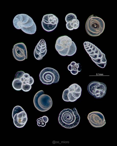
Foraminifera, single-celled marine plankton, come in a variety of shapes and sizes. These samples were found on the beach of Sagami Bay, Japan, and arranged into this stunning slide by the microscopist.
Image courtesy of @co_micro. Captured using an Olympus BH-2 microscope.
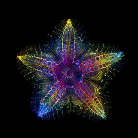
Once again, this stunning image of the nervous system of a juvenile sea star (Patiria miniata) earns a spot in our top images roundup. Fun fact: this image is also the shining star of our Image of the Year 2022 competition, winning the global award!
“This image shows the nervous system of a sea star (Patiria miniata). The main feature of the nervous system in sea stars are three long tracts of neurons called radial nerve cords, which run on the oral side along the midline of each of the five arms. These five nerve cords are linked together by a nerve ring that circles around the mouth. From the nerve cords, regularly spaced lateral nerves branch toward the edges of each arm.”
Image and quote courtesy of Laurent Formery, global winner of the 2022 Image of the Year competition. To see all the winning images, visit olympus-lifescience.com/ioty.
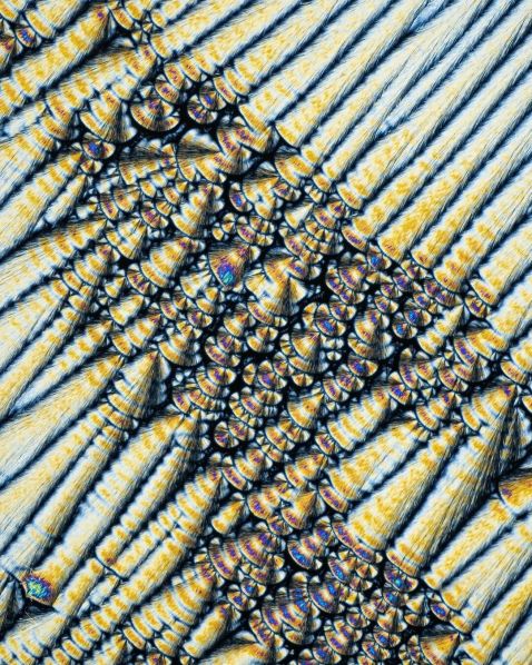
With the winter weather and busy holiday season, we all need to take care of our immune systems. This image shows us how beautiful vitamin C can look—it’s healthy and pretty!
"Here's a frame I created a few weeks back of vitamin C crystals on a glass slide. The frame was created through the phenomenon of birefringence using a 20X objective. Despite the minuscule scale of these crystals, they reveal intricate details and stunning patterns that can only be appreciated through the lens of a microscope."
Image and caption courtesy of Raghuram Annadana. Captured using an Olympus CX43 microscope.
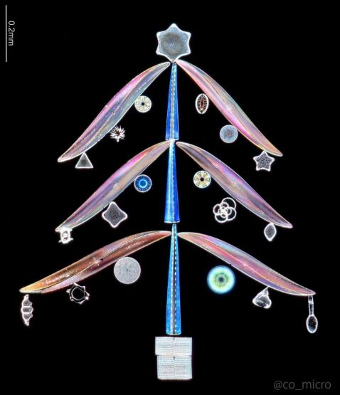
Our friends the foraminifera make a second appearance this month—this time with their fellow microorganisms diatoms, radiolarians, and sponge spicules. These specimens were collected in Sagami Bay and the Izu Islands, Japan. Again, the microscopist arranged them on a slide before imaging them, but this time created a festive tree, holiday ornaments and all!
Image courtesy of @co_micro. Captured using an Olympus BH-2 microscope.
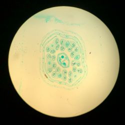


Rapeseed oil, often know as canola oil, is used for cooking due to its high smoke point (among other reasons). It’s cultivated from from rapeseed, a bright yellow flower. These rapeseed flower sections are smiling at us and made us smile too!
Image courtesy of UiM. Captured using an Olympus BH2 microscope.
As a bonus, check out this video of white blood cells to the rescue!
"To simulate an infection, I added yeasts and green algae to a drop of my blood.
Instantly, white blood cells (most probably macrophages and neutrophils) were attracted by the fake pathogens I introduced them! Like little amoebas, they crawled up to foreign organisms before eating and digesting them, a process called phagocytosis. This way, the danger is neutralized and won’t be able to hurt you!
The role of your immune system is to protect you against a wide array of pathogens like viruses, bacteria, fungi, and parasites like worms or protozoa. It seems straightforward, but it’s a bit more complicated than this!
There are two different types of immune responses against invading microbes: the innate response and the adaptative response. They both work together to eliminate a threat but are very different responses. In this video, you can observe the innate response, also called natural response, in action! This response is the body’s first line of defense against pathogens and is rapid and non-specific. This means it can recognize a wide range of pathogens that share common features and uses phagocytic cells like neutrophils and macrophages to destroy them. If you get bitten by an insect, macrophages and neutrophils will run at the crime scene and neutralize toxins or pathogens, if some got in. If there’s an infection and pathogens couldn’t be destroyed by macrophages and neutrophils, lymphocytes come along to the party!"
Video and caption courtesy of Chloé Savard. Captured using an Olympus BX53 microscope.
To see more images like these, be sure to follow us on Instagram at @evidentlifescience!
Interested in sharing your own images? Visit our image submission site!
Related Content
Over the Rainbow—Our Most Popular Microscope Images for November 2023
Pretty Pollen—Our Most Popular Microscope Images for October 2023
Magnificent Microorganisms—Our Most Popular Microscope Images for September 2023
.jpg?rev=2CBF)
