The top microscope images from November boast a multitude of bright colors. While we are used to seeing certain colors in objects, viewing them under the microscope (and using various stains) often enables us to see specimens in a whole new way. From moss piglets to pine, the microscopic world is a technicolor one.

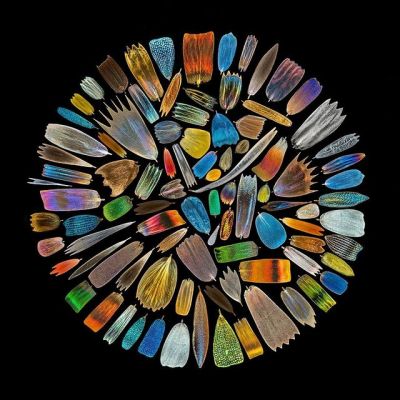
For the second month in a row, the most-liked image was a winning image from our Global Image of the Year (IOTY) competition. This image is our 2020 IOTY regional winner for Asia-Pacific. Scales were collected from the wings of more than 40 species of butterflies, photographed individually, and assembled to form this stunning microscopic mandala.
To see the rest of our winners, or to submit your own image to our 2022 competition, visit our IOTY page.
Image courtesy of XinPei Zhang, 2020 IOTY regional winner for Asia-Pacific. Captured using an Olympus Vanox AH microscope.

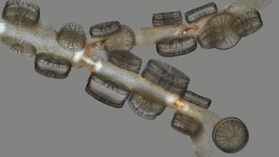
This specimen almost looks good enough to eat! While they may look like Oreo cookies, these are actually Arachnoidiscus japonicus diatoms adhered to seaweed, in situ. This sample shows various angles of the diatom. We may not want to dip them into milk as a snack, but they likely look delicious to other organisms. Diatoms can photosynthesize, so they convert dissolved carbon dioxide in water into oxygen. This makes them a primary food source for invertebrates and small fish!
Image courtesy of Macro Cosmos Microscopy. Captured using brightfield with extensive Z-stacking on an Olympus AX70 microscope.


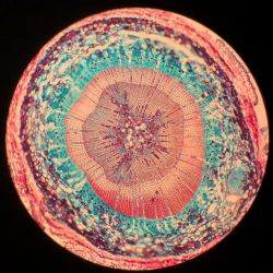
Commonly found in the Northern Hemisphere, a pine is any conifer tree or shrub in the genus Pinus of the family Pinaceae. The term “pine” may also refer to the wood from these trees, as it is one of the most frequently used types of lumber. When we think of pine trees, we usually think of the color evergreen. These stem cross sections instead make us think of peach, purple, and teal!
Image courtesy of UiM. Captured using an Olympus BH2 microscope.


Many of you correctly put together this puzzle and immediately recognized this beautiful fluorescence image. Our 2019 IOTY regional winner for the Americas shows a tardigrade (also called a water bear or moss piglet) stained to show the colorful details of its insides. While we all love moss piglets for their microscopic cuteness and resilience, this stunning multicolored image makes our favorite microorganism look ready to party!
Image courtesy of Tagide deCarvalho, 2019 IOTY regional winner for the Americas.

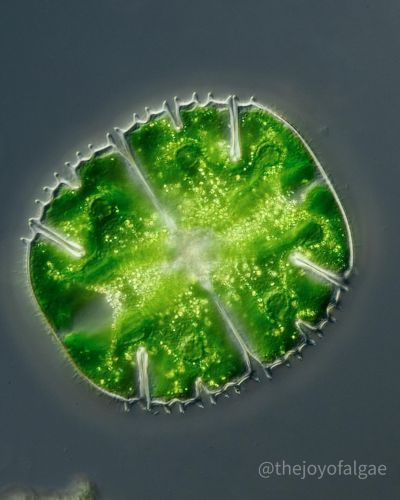
“This tiny sparkling green alga is Micrasterias truncata, collected from a wild place where land meets the sea at Lower Bostraze. Close to the home and a known sampling site of influential botanist John Ralfs (1807–1890), it's the most southerly bog on the UK mainland. Along with beautiful and accurate illustrations by Edward Genner, Ralfs published The British Desmidieæ in 1848. The plate displaying M. truncata shows it as a juvenile form of its larger relative M. rotata. Ralfs corrects this in the text following correspondence with eminent French botanist Louis Alphonse de Brébisson, who first described the species in 1846. I like to think this might even be an ancestor of the very M. truncata strain observed by Ralfs all those years ago (it’s entirely possible).”
Image and caption courtesy of Dan, the Joy of Algae. Captured using an Olympus BX51 microscope at 100X magnification.
To see more images like these, be sure to follow us on Instagram at @olympuslifescience!
Interested in sharing your own images? Visit our image submission site, and be sure to enter your best light microscopy images into our 2022 Global Image of the Year contest.
Related Content
From Tinkerer to Prize Winner: Meet the 2021 Recipient of the IOTY Award for Asia
Creepy Critters―Our Most Popular Microscope Images for October 2022
Celebrating the Global Image of the Year EMEA Winner’s Festive Fungi
.jpg?rev=6F97)
