As scientists look for new insights about how the mind and body works, they will need to dig deeper than ever before. Multiphoton microscopy is becoming a popular technique to do just that. This powerful imaging method can noninvasively capture 3D images of dynamic cellular events deep in live tissue.
Yet researchers often overlook an optical challenge that occurs during multiphoton deep imaging—spherical aberration.
Here, we take an in-depth look at spherical aberration in multiphoton deep imaging and discuss how to overcome it.
What is spherical aberration?
Simply put, spherical aberration is an optical error that occurs when light rays pass through a spherical lens and converge at different points. Since the rays fail to meet at one focal point, image brightness and resolution suffer. In deep brain regions, for example, spherical aberration makes it difficult to study fine structures like dendritic spines.
Fortunately, innovations in microscope objective technology can compensate for this error. Scientists can use correction collars on objectives to shift the position of internal lens elements and fix spherical aberration. Yet, fixing spherical aberration with a manual correction collar during deep imaging is anything but simple.
Here are two common problems:
- The darkroom environment of multiphoton systems makes it difficult to see and adjust the collar position
- Each collar adjustment slightly alters the effective focal length of the objective
With these challenges, it becomes impractical to manually adjust a collar for more than a single position during volume acquisition of Z-stack images—which can limit your ability to capture bright and high-resolution images at every depth.
To overcome this difficulty, we recommend using a motorized lens system like our TruResolution objectives. Here are two key benefits:
1. Simplify user operation.
Multiphoton microscopy is an advanced fluorescence imaging technique, so some scientists may not feel confident acquiring images without a microscope expert present. Motorized objectives can greatly simplify user operation for multiphoton deep imaging experiments by automating spherical aberration correction. Here’s how it works:
As you can see in the image below, the focal plane changes when conventional collars are rotated (Figure A, left). In comparison, motorized objectives automatically change the Z-position of an objective according to the rotation angle. It also optimizes the collar based on objectively measured quantities, such as image contrast (Figure B, right).

With this innovative technology, the software control simplifies user operation under a range of external conditions.
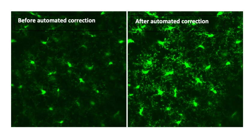
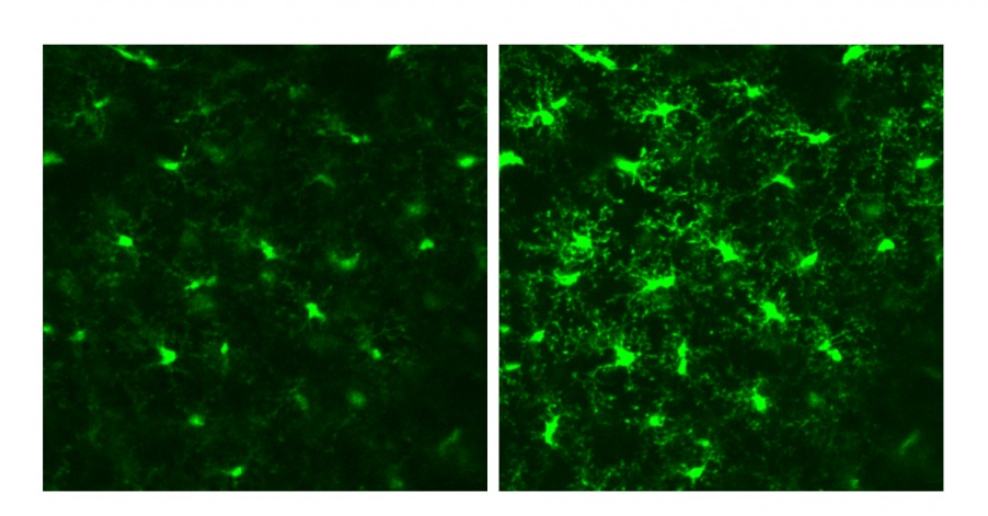
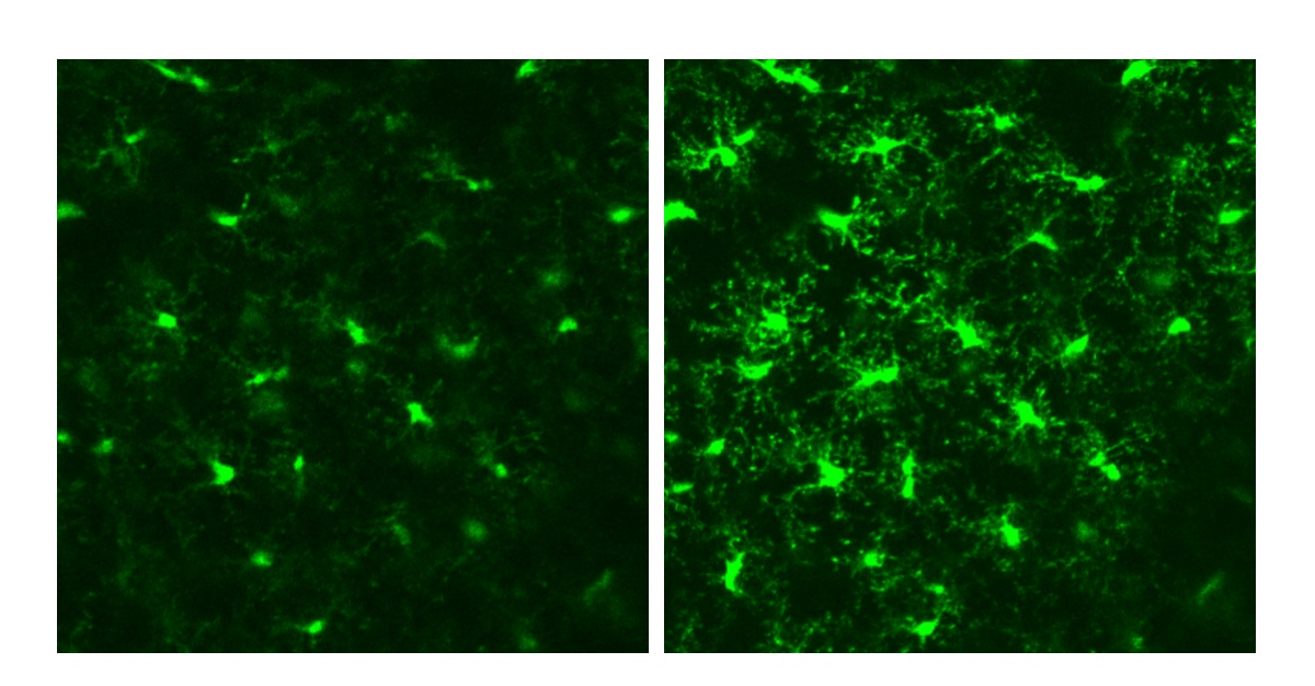
You can see the difference in the two images below of microglia 100 μm deep in a live mouse visual cortex. The image acquired after automated collar correction (right) is brighter and better resolves fine filopodia-like protrusions compared to the image captured prior to collar adjustment (left).

Images courtesy of Mitchell Murdock of Massachusetts Institute of Technology (MIT).
2. Provide bright, clear images at every depth.
An automated correction collar can tailor optical corrections to depth and refractive index profiles, helping you capture brighter images and finer features deep inside biological tissue.
For example, neuroscientists are interested in the structural morphology of submicron features, such as dendritic spine heads and necks, through deep regions in the brain. With clearer and sharper images, they can better characterize these dendritic spines to study learning and memory.
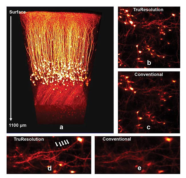
Here’s what this looks like in practice. In a recent study, researchers performed in vivo observation of the sensory cortex in an anesthetized mouse prepared with a glass cranial window.

Images courtesy of Hiromu Monai, Hajime Hirase, and Atsushi Miyawaki at RIKEN BSI-Olympus.
The image captured with an automated TruResolution objective (top right, B) features higher resolution and brightness compared to the one acquired with the correction collar fixed at the surface (middle right, C).
The improved image quality from the TruResolution objective makes the dendritic spine details easier to observe (bottom left, D) than in the image captured with the fixed correction collar (bottom right, E).
Make Deeper Discoveries
Multiphoton imaging at deep levels can help researchers better understand neurological diseases and disorders, such as Alzheimer’s, Parkinson’s, and multiple sclerosis. Our motorized objectives for the FVMPE-RS multiphoton laser scanning microscope can provide the clear, bright, and precise images you need to zero in on details and find the next life-changing discovery.
Related Content
Video: TruResolution: Maximize Resolution in Deep Imaging
Webinar: Multiphoton Microscopy for Deep Tissue Penetration
White Paper: TruResolution Objectives Maximize Resolution in Deep Imaging


