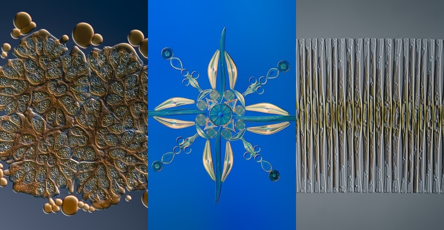
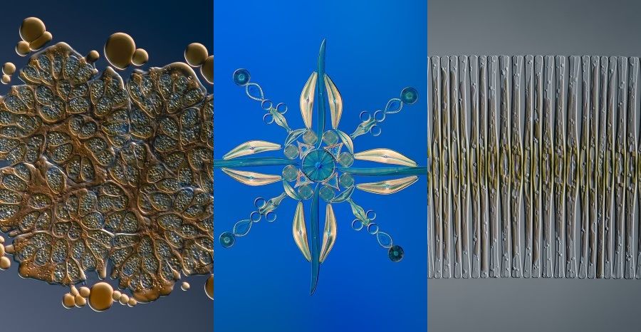
Håkan Kvarnström is a microscopy enthusiast who has been using our imaging systems and objectives to create stunning microscope artwork for many years (you can find many examples by going to his Instagram page). We first spoke to Håkan after he won first prize in our 2018 Image of the Year Award with a truly magnificent image of a sea snail.

The winning image of our 2018 Image of the Year Award. Håkan captured a 2 mm snail found at the bottom of a water bottle.
Recently, Håkan has been imaging some of his favorite subjects—pond-dwelling algae and diatoms—with our X Line objectives.
We launched this range of barrier-breaking objectives for life science in 2019 to address a longstanding tradeoff between three critical parameters—numerical aperture (NA), image flatness, and chromatic correction. An improvement in one parameter typically compromised another—but X Line optics shattered those optical barriers by improving all three areas at once.
The ability to simultaneously improve these three key parameters makes X Line objectives highly beneficial for a wide range of applications and users, such as researchers and clinicians. But they can also be used to create beautiful microscopy art.
Now, Håkan has kindly offered to share some of his X Line images with us, showing the microscopic life of the ponds in and around the royal gardens in Stockholm, Sweden. Take a look!
1. Botryococcus braunii
First up is the Botryococcus braunii: a microalga notable for its copious oil production. In fact, in this image you can see droplets of oil around the edges that were squeezed out of the specimen as water evaporated and the coverslip began to press down on the specimen.
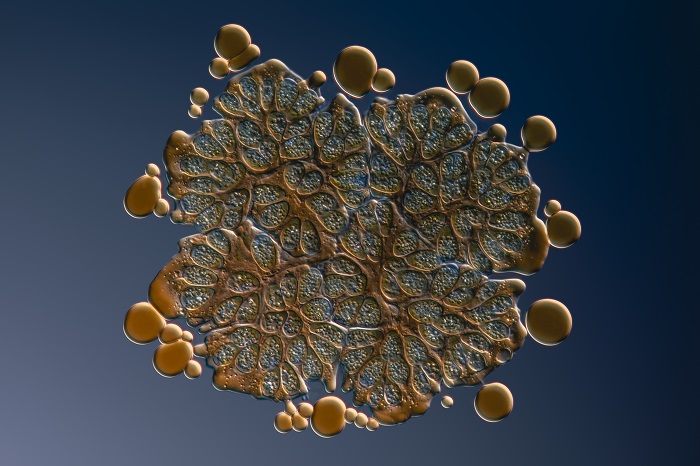
Botryococcus braunii: focus-stacked image taken with the 60x oil immersion X Line objective (UPLXAPO60XO) with a 1.42 NA. The false-coloring effect was created through deliberate misalignment of the differential interference contrast (DIC) setup.
Håkan explained how X Line optics helped him achieve this detailed image.
“The 60x X Line objective gave me the ability to resolve details that were hard to see with my other objectives. Its working distance is also considerably higher, so I can capture thicker specimens without losing sharpness and resolution,” said Håkan.
2. Fragilaria crotonensis
The same 60x objective also produced the next image of an impressive-looking freshwater diatom: Fragilaria crotonensis.
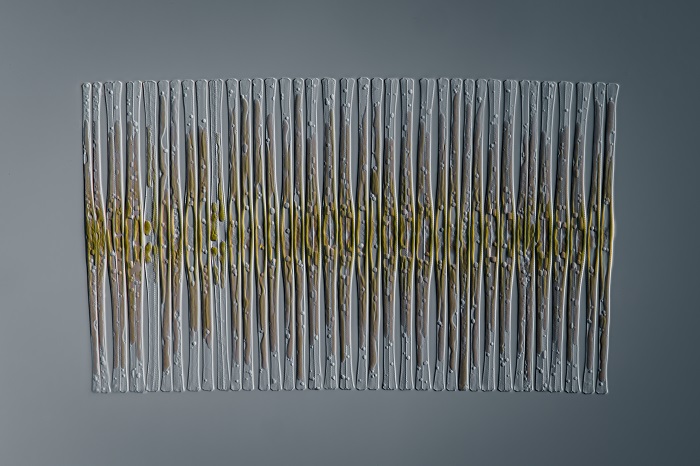
Fragilaria crotonensis: focus-stacked DIC image (60x oil immersion X Line objective).
Håkan commented, “The combination of 60x magnification and the high numerical aperture of 1.42 allowed me to capture the whole specimen in one frame at high resolution. My previous 100x objective had an NA of 1.40, so with X Line I get more into a frame and see more details.”
3. Diatom arrangement
The other objectives in our X Line series also offer improved specifications such as higher NA. For instance, the diatom image below was captured using a 20x objective with a 0.8 NA.
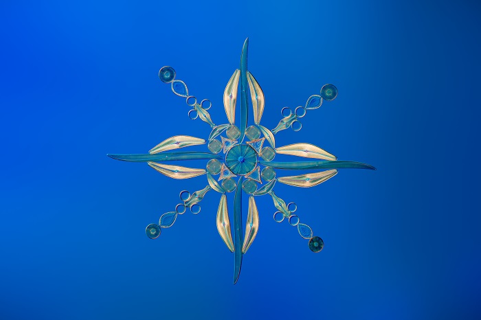
Diatom arrangement: focus-stacked DIC image (20x dry X Line objective, UPLXAPO20X).
Håkan said, “The X Line 20x objective was my absolute favorite in terms of image quality, clarity, and brightness. The increased NA going from my previous 20x objective (0.75) to X Line (0.8) has a clear advantage in that I can capture more details of the diatoms’ striae.”
4. Vorticella
The final image of this venture into the world of X Line optics is this single-shot image of a live, fast-moving Vorticella.
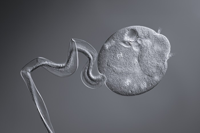
Vorticella: DIC image (40x dry X Line objective, UPLXAPO40X).
It highlights the benefit of good chromatic correction.
Håkan explained, “Contrary to what many believe, this correction is also important in monochrome images. Chromatic aberration converted to black and white gives a softness to edges, which is something I always try to avoid, especially where sharp edges are a characteristic feature. The 40x X Line objective produced excellent sharpness with no visible aberrations.”
This image also highlights why a high working distance is so important.
“High working distance is really important for living specimens because they move around and you have to give them space,” said Håkan. “Specimens like this Vorticella don’t just move back and forth. They also move up and down—in and out of focus. So, the higher the working distance, the better.”
The images here are just a small sample of what’s possible with X Line objectives. To see more, you can follow Håkan on Instagram and be sure to give Olympus Life Science a follow as well.
Curious to see how X Line objectives can help your work? Learn more about our high-performance microscope objectives here.
Related Content
2018 Image of the Year Award Spotlight: 1st Prize Winner Håkan Kvarnström Captures the Golden Ratio
Revealing the Beauty of the Microscopic Scale—IOTY’s 2019 Asia-Pacific Regional Winner
