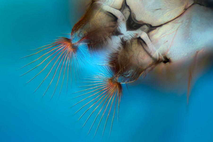
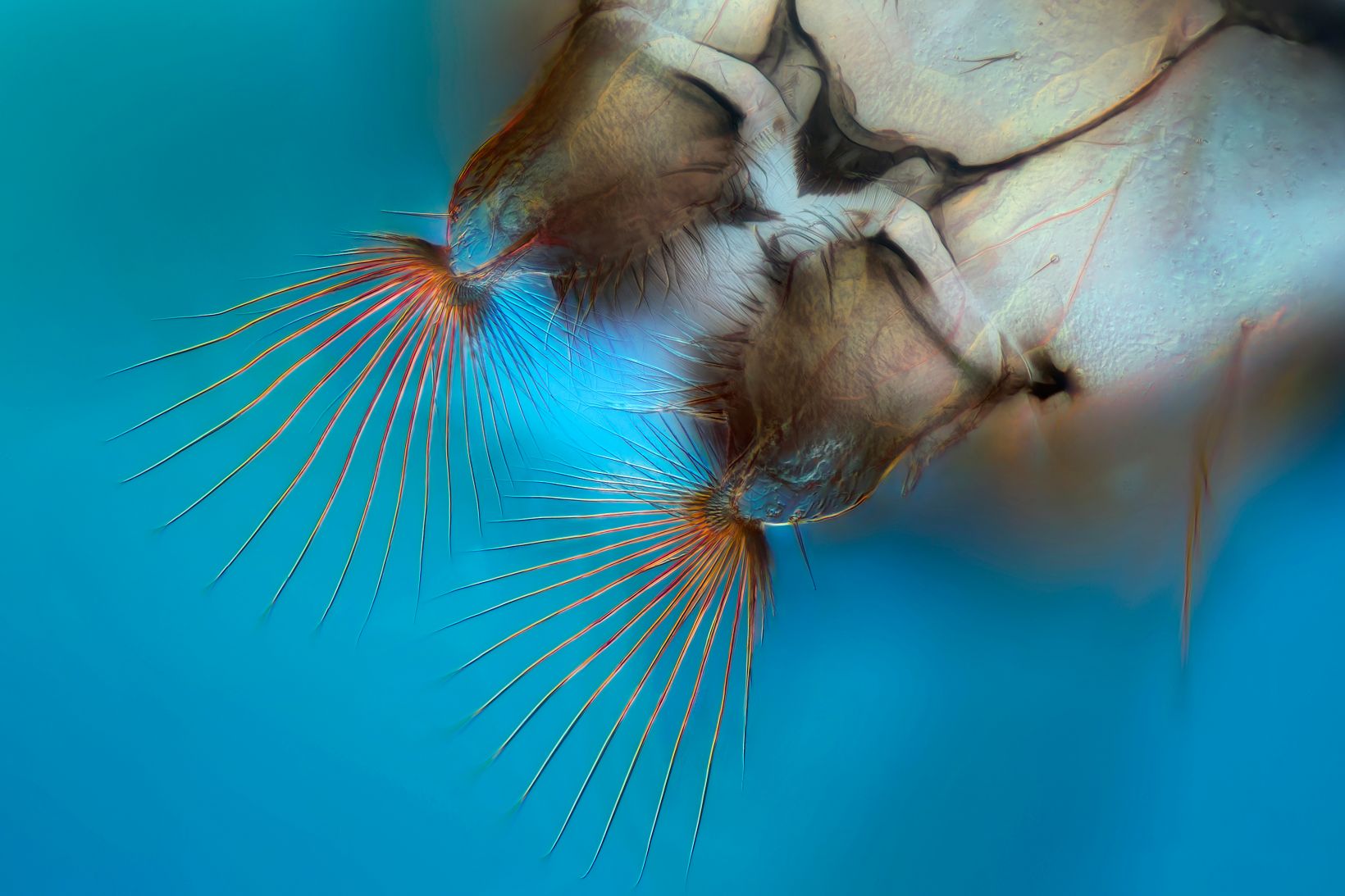
Is it an alien lifeform? Creature from the deep? No, the fascinating image below captured by Johann Swanepoel is actually a humble mosquito larva showing off its distinctive mouth brushes.
This image won third prize in our 2018 Image of the Year (IOTY) European Life Science Light Microscopy Award, our competition that recognizes the best in life science imaging.
While we received many incredible submissions, our judges were impressed by the level of detail captured in this image.
We asked Johann how he did it.
He explained that he captured the image at 400x magnification with an Olympus BX53 microscope using contrast methods and image stacking.
“I used my Olympus BX53 microscope with differential interference contrast (DIC) and a camera. DIC uses prisms to give you better contrast, which is particularly helpful when your samples are transparent,” Johann elaborated.
When capturing images at 400x magnification, the depth of field is very low, meaning that only a small part of a 3D object like this will be in focus.
With that in mind, how did Johann get such a perfectly focused image?
Here’s the secret: The image is composed of several photographs, all focused at different depths, stacked together using software to form the final image.
“This image consists of 16 images stacked together,” Johann explained. “You can either stitch the images taken at different depths together manually or use specialized software. I used a software package.”
For Johann, microscopy is a hobby that combines all his passions.
“I build websites and I do a lot of coding and use image software in my day-to-day job. It’s great to be able to combine that with microbiology and photography through the image stacking — I haven't got bored of it in the last three years!”
So, how does he feel about winning an Image of the Year Award?
“I think the IOTY competition was a great way to get recognized for something that brings a lot of joy to my life. I’ve always really liked that image, it's my personal favorite, so it’s really great to win an award for it!” Johann said.
As we celebrate this award-winning image, our search for the best light microscopy imaging continues—this time on a global scale. This year we’re proud to announce Olympus’ first Image of the Year Global Life Science Light Microscopy Award.
To learn more about the IOTY Award 2019, including the global and regional prizes, full terms and conditions, and jury members, visit Olympus-LifeScience.com/IOTY. And while you’re there, be sure to read Johann’s full story, download his image as inspiration, and submit your own light microscopy masterpieces.
“I’d definitely recommend entering the competition” Johann said. “It’s easy to do, and it’s great that your image gets shared and talked about. The hardest part is choosing which images to enter!”


