Judges 2009

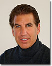
 | Mr. Thomas Deerinck - Thomas Deerinck is a research scientist at the National Center for Microscopy and Imaging Research (NCMIR) at the University of California, San Diego. He specializes in developing biological specimen labeling techniques for many types of microscopies including confocal, multiphoton and electron microscopy as well as electron tomography. He has won numerous awards including first place in the 2006 Olympus BioScapes Competition and the Crowley Award for outstanding contributions to the field of electron microscopy from the Microscopy Society of America in 2008. His work has appeared in such prestigious scientific journals as Nature, Science, and Cell and his images have been featured in various periodicals such as National Geographic, Scientific American, Discover and Time. He is currently working with teams headed by Mark Ellisman, director of NCMIR, and Roger Tsien, winner of the 2008 Nobel Prize in Chemistry, in advancing methods for correlated light and electron microscopic imaging. |
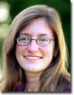
 | Dr. Julie Theriot - Dr. Theriot is Associate Professor in the Departments of Biochemistry and of Microbiology & Immunology at Stanford University School of Medicine, Stanford, CA. She has worked extensively in the fields of cell motility and locomotion, actin dynamics and intracellular pathogens. She holds two BS degrees (physics and biology) from the Massachusetts Institute of Technology, Cambridge, MA, and a PhD from the University of California, San Francisco in cell biology. A recognized expert in photomicrography, she has captured many images that have appeared in cell biology and microbiology textbooks. She has earned numerous honors in her teaching and research career, including a John D. and Catherine T. MacArthur Foundation Fellowship and the Stanford University School of Medicine Award for Graduate Teaching; she has also been named a Howard Hughes Medical Institute Investigator. She has authored scores of papers, invited reviews and book chapters, and is an author of the book Physical Biology of the Cell; in addition, she has served on several prestigious National Research Council committees and sits on a number of publication editorial boards. |
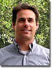
 | Dr. Kenneth N. Fish - An Assistant Professor in the Translational Neuroscience Program in the Department of Psychiatry at the University of Pittsburgh, Dr. Fish uses knockout and genetic mutants that have subtle neuroachitectural abnormalities to study the functional consequences that mispositioning of a subset of neurons during development has on local circuitry. Studies using these mutants may be helpful in understanding aspects of the neuropathology that result in abnormal brain connectivity associated with schizophrenia, autism, and epilepsies. Optical microscopy techniques utilized in this research include confocal and multiphoton fluorescence imaging, as well as total internal reflection fluorescence (TIRFM) along with traditional widefield fluorescence methods. Dr. Fish also has experience with brightfield imaging modes, including phase contrast, differential interference contrast, and oblique illumination. |
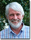
 | Dr. Douglas B. Murphy - Professor Murphy is the Director of the optical and electron microscopy laboratory at the Howard Hughes Medical Institute in Janelia Farm, Virginia. This resource is a core facility that provides equipment and services in fluorescence, confocal, and electron microscopy to laboratories throughout the research campus. Dr. Murphy's research interests include the dependence of microtubules in organelle transport and related microtubule behavior, including polymer annealing and the mechanism of binding microtubule-associated protein (MAP-2) on microtubule surfaces. He has also explored the mechanical and conformational requirements of the motor protein, kinesin. The core facility specializes in advanced fluorescence techniques (FRET, FRAP, TIRFM), confocal, multiphoton, and time-lapse in multiple fluorescence channels. Dr. Murphy has been a significant contributor to review articles and Java tutorials on the Olympus optical microscopy educational websites. |
Not Available in Your Country
Sorry, this page is not
available in your country.