Evident LogoOlympus Logo

Ask the Experts

Olympus Discovery Summit: Advance Your Imaging | October 26–27, 2021

Olympus Discovery Summit: Advance Your Imaging

Register Now


Agenda

Tuesday, October 26, 2021 Wednesday, October 27, 2021

12:00pm - 12:50pm

6:00am - 6:50am

12:00am - 12:50am

The Use of Multiplexing in Microscopy for Better Understanding the Skin Immune System in the Context of the Tissue

Presenter:
Alfonso J. Schmidt

Senior Staff Scientist
Malaghan Institute of Medical Research
 

More information

Evolution of Scientific Digital Imaging Technologies and their Applications

Presenter:
Guo Lin, Ph.D.

Manager, Product & Applications
Olympus Singapore
 

More information

1:00pm - 1:50pm

7:00am - 7:50am

1:00am - 1:50am

Now You Have the Power to See More

Presenter:
Sandrine Roy, Ph.D.

Business Development Manager
Olympus Australia & New Zealand
 

More information

Hyperspectral and Brightfield Imaging Combined with Deep Learning Uncover Hidden Regularities of Colors and Patterns in Cells and Tissues

Presenter:
Prof. Ewa Goldys, Ph.D.

Deputy Director
Australian Research Council Centre of Excellence in Nanoscale Biophotonics
 

More information

2:00pm - 2:50pm

8:00am - 8:50am

2:00am - 2:50am

A Live Demonstration of the SLIDEVIEW™ VS200 Research Slide Scanner

Demonstrators:
Chunsong Yan

Business Development Manager
Olympus Australia & New Zealand
Tong Wu, Ph.D.
Business Development Manager
Olympus Australia New Zealand
 

More information

A New Way of Thinking—Object Detection with Deep Learning

Presenter:
Akira Saito

Assistant Manager, Marketing and Applications
Olympus Singapore
 

More information

3:00pm - 3:50pm

9:00am - 9:50am

3:00am - 3:50am

Recent Advances in 3D Imaging and AI-Driven Data Analysis

Presenter:
Seungil Kim, Ph.D.

Staff Scientist
Ellison Institute for Transformative Medicine at the University of Southern California
 

More information

Confocal Microscopy and Its Use for a Spaceflight Experiment

Presenter:
RJC (Bob) McLean, Ph.D.

Regents’ Professor
Texas State University
 

More information

4:00pm - 4:50pm

10:00am - 10:50am

4:00am - 4:50am

Deep Learning Approaches to Automated Phenotypic Profiling

Presenter:
Jesse Chao, Ph.D.

Scientist
Sunnybrook Research Institute
 

More information

Live Demo: SLIDEVIEW™ VS200 Research Slide Scanner

Demonstrators:
Wei Juan Wong

Application Specialist
Olympus Soft Imaging Solutions
 

More information

5:00pm - 5:50pm

11:00am - 11:50am

5:00am - 5:50am

Accelerating Image Analysis with TruAI™ Deep Learning Technology

Presenter:
Manoel Veiga, Ph.D.

Application Specialist
Olympus Soft Imaging Solutions
 

More information

The Use of Multiplexing in Microscopy for Better Understanding the Skin Immune System in the Context of the Tissue

Presenter:
Alfonso J. Schmidt

Senior Staff Scientist
Malaghan Institute of Medical Research
 

More information

6:00pm - 6:50pm

12:00pm - 12:50pm

6:00am - 6:50am

Break

7:00pm - 7:50pm

1:00pm - 1:50pm

7:00am - 7:50am

Metabolic Imaging in Langerhans Human Islets with MPE and FLIM

Presenter:
Francesco Cardarelli

Associate Professor in Applied Physics
Scuola Normale Superiore of Pisa
 

More information

In-Vivo Tracking of Harmonic Nanoparticles by Means of a TIGER Widefield Microscope

Presenter:
Laura Vittadello, Ph.D.

Department of Physics and Center of Cellular Nanoanalytics
Osnabrück University
 

More information

8:00pm - 8:50pm

2:00pm - 2:50pm

8:00am - 8:50am

Live Demo: IXplore™ SpinSR Confocal Super Resolution System

Demonstrator:
Stefan Marawske

Senior Sales Specialist, High-End Life Science Systems
Olympus Europa
 

More information

Live Demo: FLUOVIEW™ FV3000 Confocal Laser Scanning Microscope

Demonstrator:
Bülent Peker

Senior Product Marketing Manager
Olympus Europa
 

More information

9:00pm - 9:50pm

3:00pm - 3:50pm

9:00am - 9:50am

Deconvolution of 3D Image Stacks

Presenter:
Heiko Gäthje

Senior Trainer
Olympus Europa
 

More information

Whole-Brain Functional Calcium Imaging Using Light Sheet Microscopy

Presenter:
Ruben Portugues, Ph.D.

Institute of Neuroscience
Technical University of Munich
 

More information

10:00pm - 10:50pm

4:00pm - 4:50pm

10:00am - 10:50am

Human Pluripotent Stem Cell-Derived Liver Organoid Manufacturing

Presenter:
Yosuke Yoneyama, Ph.D.

Assistant Professor
Institute of Research, Tokyo Medical and Dental University
 

More information

Now You Have the Power to See More

Presenter:
Sandrine Roy, Ph.D.

Business Development Manager
Olympus Australia & New Zealand
 

More information

11:00pm - 11:50pm

5:00pm - 5:50pm

11:00am - 11:50am

Confocal Microscopy and Its Use for a Spaceflight Experiment

Presenter:
RJC (Bob) McLean, Ph.D.

Regents’ Professor
Texas State University
 

More information

Metabolic Imaging in Langerhans Human Islets with MPE and FLIM

Presenter:
Francesco Cardarelli

Associate Professor in Applied Physics
Scuola Normale Superiore of Pisa
 

More information

12:00am - 12:50am

6:00pm - 6:50pm

12:00pm - 12:50pm

Break

1:00am - 1:50am

7:00pm - 7:50pm

1:00pm - 1:50pm

Accelerating Image Analysis with TruAI™ Deep Learning Technology

Presenter:
Manoel Veiga, Ph.D.

Application Specialist
Olympus Soft Imaging Solutions
 

More information

In-Vivo Tracking of Harmonic Nanoparticles by Means of a TIGER Widefield Microscope

Presenter:
Laura Vittadello, Ph.D.

Department of Physics and Center of Cellular Nanoanalytics
Osnabrück University
 

More information

2:00am - 2:50am

8:00pm - 8:50pm

2:00pm - 2:50pm

Recent Advances in 3D Imaging and AI-Driven Data Analysis

Presenter:
Seungil Kim, Ph.D.

Staff Scientist
Ellison Institute for Transformative Medicine at the University of Southern California
 

More information

Deep Learning Approaches to Automated Phenotypic Profiling

Presenter:
Jesse Chao, Ph.D.

Scientist
Sunnybrook Research Institute
 

More information

3:00am - 3:50am

9:00pm - 9:50pm

3:00pm - 3:50pm

Live Demo: FLUOVIEW™ FV3000 Confocal Laser Scanning Microscope

Demonstrator:
James Lopez, Ph.D.

National Applications Manager
Olympus Life Science
 

More information

Hyperspectral and Brightfield Imaging Combined with Deep Learning Uncover Hidden Regularities of Colors and Patterns in Cells and Tissues

Presenter:
Prof. Ewa Goldys, Ph.D.

Deputy Director
Australian Research Council Centre of Excellence in Nanoscale Biophotonics
 

More information

Click here to register


Speakers

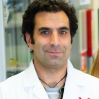
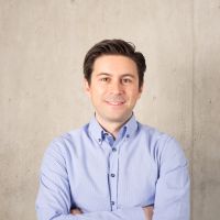
Alfonso J. Schmidt

Senior Staff Scientist
Malaghan Institute of Medical Research

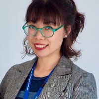
Sandrine Roy, Ph.D.

Business Development Manager
Olympus Australia & New Zealand

Alfonso J. Schmidt

Biography

Alfonso has a decade of experience working in a shared resource lab (SRL) with a vast knowledge in histology, fluorescent microscopy, and image analysis. His work has been focused in maximizing the capabilities of the equipment available and in creating technical protocols and training modules for the scientific community. Currently, Alfonso oversees the Histology and Bioimaging Facility as part of the Hugh Green Cytometry Centre (HGCC) at the Malaghan Institute of Medical Research in Wellington, New Zealand.

Abstract

The Use of Multiplexing in Microscopy for Better Understanding the Skin Immune System in the Context of the Tissue

The skin is the first line of defense and the immune system’s biggest barrier for combating pathogens. Being able to accurately characterize and identify immune cell subtypes, tissues structures, and cell distribution in the skin under steady-state conditions provides a powerful tool for understanding the first immunological strategies and biological processes that occur in the presence of pathogens. In this webinar we will review technical aspects involved in the experimental process and explore how complementary imaging technologies might assist us to better understand the immune system.

The presentation is divided into three parts. First, an introduction of the Hugh Green Cytometry Centre will be presented and an overview of the histology and bioimaging technological platforms available. Second, the multiplexing methodology will be discussed, where several topics need to be considered for the design and development of a successful polychromatic panel for microscopy. Finally, preliminary results from a research project will be presented that constitutes part of a diploma program from The Royal Microscopical Society. The project focuses on the identification of immune cell types in the whole mount skin in relation to tissues structures (e.g., blood vessels and lymphatic network). It also centers on the immune cells’ distribution in the tissue as a first barrier of defense against pathogens.

Sandrine Roy

Biography

Dr. Roy completed a double-major in Biochemistry and Microbiology followed by the completion of a Doctor of Philosophy in 2002 in the field of Molecular Biology/Cell Biology at the University of Queensland Australia. She travelled abroad to undertake a post-doctoral position at Washington University in St Louis, USA. She then returned to Australia to continue her post-doctoral studies.

With her extensive microscopy experience, she was appointed as the University of Queensland Diamantina Institute Microscopy Facility Manager in 2009 and later as manager of microscopy services at the Translational Research Institute in Brisbane until 2019.

She is now Business Development Manager at Olympus Australia, where her experience and knowledge is used to support customers both in Australia and New Zealand.

Abstract

Now You Have the Power to See More

The Olympus VS200 digital slide scanner has been very well received since its release in March 2020. With a reliable, flexible, and customizable design, the system has been adopted by various industries including research, geology, and many others. Join us to find out more and see examples of samples scanned using this popular addition to the Olympus product range.


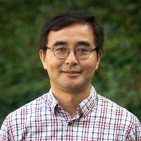
Chunsong Yan

Business Development Manager
Olympus Australia New Zealand

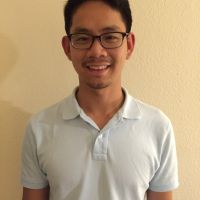
Tong Wu, Ph.D.

Business Development Manager
Olympus Australia New Zealand

Chunsong Yan

Biography

Chunsong is the business development manager for life science at Olympus Australia and New Zealand. He is currently responsible for confocal, multiphoton, light sheet, and slide scanning systems. He joined Olympus in 2003, working in various roles, always trying to offer the best Olympus solution to our customers.

Abstract

A Live Demonstration of the SLIDEVIEW™ VS200 Research Slide Scanner

Presentation Description: The Olympus SLIDEVIEW VS200 research slide scanner captures high-quality virtual slide images and enables advanced quantitative image analysis. Reliable virtual slide data can be acquired with as few as two clicks. Highly versatile, the SLIDEVIEW VS200 slide scanner supports five observation methods and a wide range of sample sizes for use in various applications. Its automatic slide loader accommodates many slide glasses, helping increase experiment efficiency.

In this session Tong Wu and Chunsong Yan will conduct a live demonstration of this product and take your through some of its benefits and features:

  1. Flexible batch scan mode that enables you to designate a different observation method such as FL, BF, POL, DF, PH for each slide contained in a batch.
  2. High-resolution fluorescence imaging using an oil immersion lens, where you will see the automatic oil dispenser in action.
  3. Live deblurring to make your images sharper and clearer during image acquisition.
  4. Data management using the NIS SQL database, which allows you to easily store, manage, and share data.

Tong Wu

Biography

Tong Wu joined Olympus in 2012 after completing her Ph.D. in China (State Key Laboratory of Fine Chemicals, DLUT). Now, Tong is a business development manager, supporting high-end microscopes in Olympus Australia. With a research background in fluorescent dyes for bio-imaging and bio-labelling, Tong Wu is enthusiastic to engage with customers’ research applications. 

Abstract

A Live Demonstration of the SLIDEVIEW™ VS200 Research Slide Scanner

Presentation Description: The Olympus SLIDEVIEW VS200 research slide scanner captures high-quality virtual slide images and enables advanced quantitative image analysis. Reliable virtual slide data can be acquired with as few as two clicks. Highly versatile, the SLIDEVIEW VS200 slide scanner supports five observation methods and a wide range of sample sizes for use in various applications. Its automatic slide loader accommodates many slide glasses, helping increase experiment efficiency.

In this session Tong Wu and Chunsong Yan will conduct a live demonstration of this product and take your through some of its benefits and features:

  1. Flexible batch scan mode that enables you to designate a different observation method such as FL, BF, POL, DF, PH for each slide contained in a batch.
  2. High-resolution fluorescence imaging using an oil immersion lens, where you will see the automatic oil dispenser in action.
  3. Live deblurring to make your images sharper and clearer during image acquisition.
  4. Data management using the NIS SQL database, which allows you to easily store, manage, and share data.

Seungil Kim, Ph.D.

Staff Scientist
Ellison Institute for Transformative Medicine at the University of Southern California

Jesse Chao, Ph.D.

Scientist
Sunnybrook Research Institute

Seungil Kim

Biography

Seungil Kim, Ph.D., is a Staff Scientist and Microscopy Team Manager at the Lawrence J. Ellison Institute for Transformative Medicine at USC. Dr. Kim completed his B.S. and M.S. degrees in South Korea. He then moved to Washington University and received a doctoral degree in Developmental Biology. He carried out his postdoctoral research in the department of Cell and Tissue Biology at UCSF. Seungil has over 10 years of experience working with various in vitro/in vivo models and advanced cellular imaging techniques. His current research focus is to understand the contributions of the tumor microenvironment to drug response, using patient-derived 3D organoids as a model system. Moreover, he is developing high-throughput automated imaging methods to screen novel drug compounds in colorectal cancer.

Abstract

Recent Advances in 3D Imaging and AI-Driven Data Analysis

This presentation will highlight various imaging techniques for 3D models, immunostaining with tissue clearing, and live imaging of organoids as well as AI-driven data analysis for high-content imaging and screening.

Jesse Chao

Biography

Jesse completed his Ph.D. at the University of British Columbia (UBC) in cell biology and genomics. He then continued his training at the University of California, San Diego. After, he switched his focus to developing machine learning approaches for assessing the physiological impacts of genetic variants associated with hereditary cancer at UBC. During this time, he started to develop deep learning approaches to automated phenotypic profiling based on high-content imaging data.

Abstract

Deep Learning Approaches to Automated Phenotypic Profiling

Quantifying cellular phenotypes is the key to all cell biology studies. However, modern imaging techniques can easily generate more data than an average user can comfortably handle. In this presentation, I will discuss two deep learning approaches, one semi-supervised and one supervised, for building image analysis pipelines. Either approach can be run on a free cloud GPU instance.


Manoel Veiga, Ph.D.

Application Specialist
Olympus Soft Imaging Solutions

Francesco Cardarelli

Associate Professor in Applied Physics
Scuola Normale Superiore of Pisa

Manoel Veiga

Biography

Manoel Veiga earned his Ph.D. in Physical Chemistry in the University Santiago de Compostela, Spain. Following two postdocs in the Universities Complutense of Madrid and WWU Münster, he joined PicoQuant GmbH. After five years supporting customers worldwide in the fields of FLIM and time-resolved spectroscopy, he joined Olympus Soft Imaging Solutions GmbH in 2017, where he works as Global Application Specialist with a focus on high-content analysis and deep learning.

Abstract

Accelerating Image Analysis with TruAI™ Deep Learning Technology

Hardware development in microscopy has enabled scientists to collect more and more image data in a very efficient and automated way. But to gain quantitative information of this data, images need to be analyzed in a robust way.

Image segmentation and classification lies at the heart of image analysis. The better the scientist can segment and classify objects of interest in their images, the better quantitative information they can gain, improving the quality of the results.

In 2019 Olympus launched TruAI technology, an image analysis approach based on deep neural networks with a focus in segmentation and classification. TruAI technology makes segmentation and classification easy and accessible by non-experts, even in the most difficult scenarios, like in low contrast brightfield images, low signal-to-noise fluorescence images, or in situations of highly dense cell confluency. When installed in the microscope that is acquiring the images, it speeds up the research as soon as analysis results can be available, shortly after the acquisition has concluded, or even at the same time as the acquisition.

In this tech talk, through a collection of examples measured with our live cell imaging systems, high-content screening station, and whole slide scanner, you will see what TruAI technology can do for your research and get a preview of what is coming next.

Francesco Cardarelli

Biography

After receiving his M.Sc. in Biological Sciences from the University of Pisa in Oct 2003 and his Diploma in Biological Sciences in the same year (both with honors) from SNS, Francesco Cardarelli worked at the NEST Laboratory of SNS as a Ph.D. student in Molecular Biophysics under the supervision of Prof. Fabio Beltram. He started his interdisciplinary research at the crossroads between cell biology and physics, using advanced fluorescence microscopy methods to study the intracellular transport properties of virus-derived peptide sequences. After graduating, he became a Post-Doctoral Fellow at the Laboratory for Fluorescence Dynamics, University of California at Irvine, under the supervision of Prof. Enrico Gratton, where he coordinated the research activity for the development of new spatial variants of fluorescence correlation spectroscopy to detect barriers to molecular diffusion/flow in live cells. In Dec 2010 he was hired by the CNI@NEST (IIT) as a Post-Doctoral Fellow. Back in Italy, he started working to develop new fluorescence-based imaging and analysis methods to study single molecules in complex biological systems with high spatiotemporal resolution. This research was boosted by a number of funded grants (and established collaborations) and by an independent scientific position, first at CNR as a Researcher, then at SNS as Professor in Applied Physics.

The focus of his research is on the development of new optical microscopy techniques to increase the amount of quantitative information that can be extracted from investigations on living matter. For instance, in recent years, he and his team introduced a number of new spatiotemporal fluctuation-analysis tools (iMSD, iRICS, nD-pCF, diffusion tensor analysis, etc.) to extract structural and dynamic properties of biological objects, from molecules to entire sub-cellular structures, in their complex natural environment. Such a toolbox is becoming a new paradigm for biophysical investigations at the nanoscale, as featured in the “New and Notable” section of Biophysical Journal (2016 Aug 23; 111(4): 677–678). In 2014, together with his Team, they demonstrated the occurrence of short-range protein Brownian motion in the cell cytoplasm, being among the first to challenge the current view of the structural organization of the crowded intracellular environment. Finally, by combining this toolbox with feedback-based orbital tracking, they demonstrated that even the nanoscopic and dynamic environment of intracellular organelles can be quantitatively probed.

Abstract

Metabolic Imaging in Langerhans Human Islets with MPE and FLIM

Capturing life (mis)regulation at the nanoscale is a crucial challenge for present and future biophysics. At this scale, the main actors are the molecules. To successfully tackle molecular behavior within living matter, optical microscopy is a valuable methodological platform: by using fluorescence as the signal, spatial and temporal details of molecular processes can be investigated quantitatively. The physiopathology of beta-cell response to glucose stimulation will be used as case study of biological/biomedical interest. The metabolic traits of beta cells will be highlighted using a straightforward combination of multiphoton microscopy, fluorescence lifetime imaging, and feedback-based orbital tracking of sub-cellular nanostructures.


Stefan Marawske

Senior Sales Specialist, High-End Life Science Systems
Olympus Europa

Heiko Gäthje

Senior Trainer
Olympus Europa

Stefan Marawske

Biography

Stefan Marawske is an expert on super resolution microscopy. During his Ph.D. in the field of physical chemistry he created a home-built microscope for localization-based super resolution and particle tracking. He was fascinated by the fact that these methods are able to break the famous Abbe limit and can resolve structures that could not be identified before. He has been working with Olympus for more than 7 years and is responsible for high-end imaging systems, such as TIRF and spinning disc.

Abstract

Live Demo: IXplore™ SpinSR Confocal Super Resolution System

In this live demo, experience the IXplore SpinSR system, designed for fast 3D super resolution imaging and prolonged cell viability in time-lapse experiments. The microscope system offers XY resolution down to 120 nm without the need for dedicated labeling procedures. Learn how to easily integrate the IXplore SpinSR microscope system into existing experiments and sample protocols to streamline your research.

Heiko Gaethje

Biography

As a biologist with a focus on neuronal development of insects and the structure of sialic acid binding neuronal proteins in mammals, Heiko Gaethje gained first experience with widefield and confocal fluorescence microscopy and image processing of 3D data.

Heiko joined Olympus in 2004 as a web content manager in the Marketing and Communication team. Since 2008, he has worked as a microscopy trainer at the Olympus Academy and is responsible for the conception and introduction of digital learning tools. In addition, he supports and conducts microscopy training courses at the EMBL Heidelberg and the Zurich Winter School on Advanced Microscopy where he regularly answers questions related to image processing and image analysis.

Abstract

Deconvolution of 3D Image Stacks

Images taken with a light microscope are never true representations of the specimen. Error sources, which have to be controlled, are sample preparation and staining protocols as well as optical aberrations and limitations of microscope and digital camera.

To turn the microscopic image into a close representation of the specimen ground truth restoration algorithms are in use. Today, we will focus on restoration of three-dimensional datasets, a process called deconvolution. The mathematics behind these methods will not be covered. Rather, the aim is to provide a brief, intuitive introduction that allows us to assess the possible uses of these filters in a specific application. For this reason, we will demonstrate these filters live on selected example images.


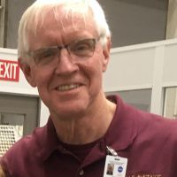
RJC (Bob) McLean, Ph.D.

Regents’ Professor
Texas State University

James Lopez, Ph.D.

National Applications Manager
Olympus America Inc.

RJC (Bob) McLean

Biography

Bob McLean has over 30 years’ experience as a microbiologist, during which time he and his lab have done a number of studies on surface-adherent microorganisms (biofilms). In 1998, he and his colleagues had an experiment on the space shuttle with John Glenn, in which they were one of the first research groups to show that biofilms could form in microgravity. Since that discovery, there have been a number of biofilm issues, notably instances of fouling in the water recovery system in the International Space Station and other spacecraft. In 2015, Bob, along with collaborators at Arizona State and the Johnson Space Center, received a NASA grant to study biofilm formation during spaceflight. Confocal and other types of microscopy have been instrumental in these investigations.

Abstract

Confocal Microscopy and Its Use for a Spaceflight Experiment

Spaceflight experiments represent a rare but exciting scientific opportunity. Unlike most lab experiments, in which protocols can be quickly modified, limitations on crew time and availability of supplies are notable factors. Unanticipated changes to launch and reentry schedules are also an issue. The experimental apparatus and protocols used must be able to function in a microgravity setting, while also resisting the g-forces and vibrations during launch and landing. During this presentation, Dr. McLean will go over the experimental planning and use of confocal and electron microscopy approaches and analyses during a recent spaceflight experiment that flew on Space X-21 from 12/6/20 – 1/14/21

James Lopez

Biography

James Lopez received his Ph.D. in biomedical sciences from the University of Chicago in 2010. With nearly a decade of experience in calcium imaging, FRET, live cell imaging, and intravital imaging, James joined Olympus as a confocal and multiphoton sales representative. He later transitioned to the Olympus Life Science Applications Group, supporting confocal and multiphoton systems. Now he manages the Life Science Applications Group in the US, Canada, and Latin America markets.

Abstract

Live demo: FLUOVIEW™ FV3000 Confocal Laser Scanning Microscope

Join James Lopez, Ph.D., National Applications Manager to see how the FV3000 confocal laser microscope can expand your research possibilities and help you get more data from your samples.


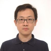
Guo Lin, Ph.D.

Manager, Product & Applications
Olympus Singapore

Prof. Ewa Goldys, Ph.D.

Deputy Director
Australian Research Council Centre of Excellence in Nanoscale Biophotonics

Guo Lin

Biography

Lin got his Ph.D. in 2010 at National University of Singapore, working on biophysical research. From 2009, he joined Olympus as Technical and Application specialist responsible for laser-based, high-end imaging systems. In 2012, Lin decided to move back to China and accept a position with a leading scientific camera manufacturer. There, he started as application specialist, later become regional sales manager, and finally scientific sales manager for Asia Pacific. In 2021, Lin moved back to Singapore, joining Olympus Singapore as the manager for products and applications. Lin has a lot of experience using various scientific digital imaging techniques, including various camera technologies.

Abstract

Evolution of Scientific Digital Imaging Technologies and their Applications

As important as an optical microscope system is to provide images with high resolution and contrast, digitizing the image seen through the eyepiece is equally important. This dramatically increases your ability to enhance features, extract information, and tell a meaningful story.

We have come a long way with respect to image capture, from photomicrography on a film to highly sensitive digital imaging that allows you to detect even single molecules. Digital imaging has allowed scientists to not only record their data, but also analyze it using new software technologies with artificial intelligence.

Digital cameras designed for life science have evolved considerably, and numerous technologies are available. From CCDs, to EMCCDs, to sCMOS, each technology has pros and cons and must be carefully chosen based on the application.

In this talk, I will cover some of the critical facts about scientific digital cameras. I will also talk about the evolution of these cameras, the solution that Olympus offers, and how they are used in current advanced microscopy systems for various applications.

Ewa Goldys

Biography

Professor Ewa M. Goldys is Deputy Director of the Australian Research Council Centre of Excellence in Nanoscale Biophotonics (cnbp.org.au) and Professor at the Graduate School of Biomedical Engineering, the University of New South Wales, Sydney, Australia.  She is Fellow of SPIE, OSA, the Australian Academy of Technological Science and Engineering (ATSE), and winner of the 2016 Australian Museum Eureka Prize for ‘Innovative Use of Technology.’ She has ongoing involvement with SPIE BIOS, the world's largest international biomedical optics meeting and SPIE's Photonics West where she serves as Track Chair in Nanobiophotonics.

Her research spans the areas of biomedical science, bioimaging, biosensing, and materials science. She developed novel approaches to biochemical and medical sensing and deployable medical diagnostics. Current projects focus on cancer nanotechnology and non-invasive high-content imaging of colors and patterns in cells and tissues.

Abstract

Hyperspectral and Brightfield Imaging Combined with Deep Learning Uncovers Hidden Regularities of Colors and Patterns in Cells and Tissues

The Australian Research Council Centre of Excellence for Nanoscale Biophotonics draws on key advances of the 21st century, nanoscience, and photonics to help understand life at the molecular level. In this presentation, next-generation technologies developed in our Centre for probing, imaging, and interacting with the living systems will be discussed. These address the key challenges of ultrasensitive detection of key analytes in real complex environments and molecular complexity, and they support both novel therapies and diagnostics.


Akira Saito

Assistant Manager, Marketing and Applications
Olympus Singapore

Yosuke Yoneyama, Ph.D.

Assistant Professor
Institute of Research, Tokyo Medical and Dental University

Akira Saito

Biography

Akira studied veterinary medicine at Tokyo University of Agriculture and Technology, Japan and graduated in 2007. Shortly after, he joined Olympus as application specialist responsible for in vivo imaging systems, high-content analysis systems, and laser confocal systems to support customers in Japan. In 2013, he took over sales promotion for all Olympus life science products. From 2018, he moved to Singapore and joined to support the marketing and application support for the APAC market.

Abstract

A New Way of Thinking—Object Detection with Deep Learning

Image analysis is widely used in life science to quantify and understand events in biological samples. Object detection and segmentation are key processes for image analysis to identify our area of interest in the images. Then we can quantify morphological information, intensities, velocities in tracking, etc.

Conventional segmentation has not always been accurate and efficient; however, our eyes and brains can identify where our areas of interest are from experience. Using deep learning, we can train a neural network with ground truth information to carry out this complex task. Once the neural network has been created properly, it can help segment objects in a similar way as your brain. Deep learning sounds like it requires programming skills, but our software does not require programming skills and is easy to use.

In this session, we will discuss object segmentation with deep learning and its applications in life science. We will also demo Olympus deep-learning software.

Yosuke Yoneyama

Biography

Yosuke Yoneyama obtained his Ph.D. from The University of Tokyo in Japan, where he continued his post-doctoral work on insulin/insulin-like growth factor signaling with a focus on spatiotemporal control of the intracellular signaling system in the laboratory of Dr. Shin-Ichiro Takahashi. He then joined the laboratory of Dr. Takanori Takebe at Tokyo Medical and Dental University in Japan as an assistant professor. He now focuses on human organoids, in particular liver organoids, that are derived from stem cells, including induced pluripotent stem cells (iPSCs), to reconstitute multiple lineages of liver cells both for human development and modeling diseases such as non-alcoholic steatohepatitis.

Abstract

Human Pluripotent Stem Cell-Derived Liver Organoid Manufacturing

Despite its translational value, organoid-based approaches are generally inefficient and irreproducible with the burden of long-term cell culture. In the case of organoids generated from human pluripotent stem cells, including induced pluripotent stem cells (iPSCs), there is a need to monitor the multiple steps that cover iPSC maintenance, two-dimensional differentiation, and three-dimensional morphogenesis. By combining a live cell monitoring system and biochemical assays, we developed a reproducible protocol for generating human liver organoids that minimizes inter-donor-dependent variabilities. Applying this setup to over 15 human iPSC libraries enabled us to investigate the genotype-phenotype relationship in liver organoids, revealing important risk gene polymorphisms for steatohepatitis and fibrosis. These results will help to build a highly reproducible organoid platform to study personalized mechanisms of health and diseases.


Wei Juan Wong

Application Specialist
Olympus Soft Imaging Solutions

Laura Vittadello, Ph.D.

Department of Physics and Center of Cellular Nanoanalytics
Osnabrück University

Wei Juan Wong

Biography

Wei Juan Wong has a degree in physics and has worked in a biophysical research laboratory as well as a microscopy core facility. She joined Olympus in 2018 as a Product Specialist at Olympus Singapore, where she supports Southeast Asia customers using widefield microscopes, including the VS200 slide scanner. In 2021, she moved to Germany to join Olympus Software Imaging Solutions as an Application Specialist, where she provides application and marketing support to customers worldwide.

Abstract

Live Demo: SLIDEVIEW™ VS200 Research Slide Scanner

In this live demo you will learn how to capture high-resolution images of your slides for quantitative analysis, enabling you to make the most of the information your slides have to offer. Easily analyze, share, and archive your data with the SLIDEVIEW VS200 digital slide scanner. Join this session and learn how to achieve more in less lead time.

Dr. Laura Vittadello

Biography

Dr. Laura Vittadello is working as a post-doc in the physics department of the Osnabrück University in the ultrafast physics research group. Her research focus is on the fundamental study and application of a new type of marker, harmonic nanoparticles, that are specially designed for biological applications that involve nonlinear microscopy.

Abstract

In-Vivo Tracking of Harmonic Nanoparticles by Means of a TIGER Widefield Microscope

In-vivo tracking based on harmonic nanoparticles is so far not exploited because of a lack of an appropriate tool—a widefield nonlinear optical microscope. Here, we present a new approach to overcome this challenge based on a redesign of laser space parameters.


Bülent Peker

Senior Product Marketing Manager
Olympus Europa

Ruben Portugues, Ph.D.

Institute of Neuroscience
Technical University of Munich

Bülent Peker

Biography

Bülent Peker is an expert on laser scanning microscopy. He first developed an interest in microscopy and photonics during his Ph.D. in physical chemistry, where he worked on time-resolved two-photon microscopy, and this passion has been with him ever since.

Bülent has been with Olympus for over 13 years and has helped the team introduce cutting-edge laser scanning microscopes to the market. He’s particularly fascinated by the application of multiphoton systems and the customization possibilities of laser scanning systems.

Abstract

Whole-Brain Functional Calcium Imaging Using Light Sheet Microscopy

Light sheet microscopy is a powerful technique to perform fast volumetric imaging. I will talk about how we use it to investigate how the brain of a small vertebrate, the larval zebrafish, works. In our laboratory at the Technical University of Munich, we are interested in how the brain processes external sensory stimuli and uses internal states and past experiences to select appropriate behavior. In order to do this, we image the activity of almost all 100,000 neurons in the brain of larval zebrafish while we present the animals with stimuli and monitor their behavior. I will also discuss the data processing steps after acquiring these large datasets.

Dr. Ruben Portugues

Biography

Prof. Portugues is a neurobiologist studying sensorimotor control. His research group uses behavior, modeling, optogenetics, in vivo electrophysiology, and whole brain functional calcium imaging to dissect learning, memory, and action selection in the larval zebrafish.

Prof. Portugues studied mathematics and did his Ph.D. in theoretical physics at Trinity College in the University of Cambridge. After a short postdoctoral fellowship in physics at Centro de Estudios Cientificos in Valdivia, Chile, he joined Professor Florian Engert’s laboratory at Harvard University and switched research interests to neuroscience. In 2014 he was appointed Max Planck Research Group Leader at the Max Planck Institute of Neurobiology in Martinsried. Since 2020 Prof. Portugues is an assistant professor at the TUM.

Abstract

Whole-Brain Functional Calcium Imaging Using Light Sheet Microscopy

Light sheet microscopy is a powerful technique to perform fast volumetric imaging. I will talk about how we use it to investigate how the brain of a small vertebrate, the larval zebrafish, works. In our laboratory at the Technical University of Munich, we are interested in how the brain processes external sensory stimuli and uses internal states and past experiences to select appropriate behavior. In order to do this, we image the activity of almost all 100,000 neurons in the brain of larval zebrafish while we present the animals with stimuli and monitor their behavior. I will also discuss the data processing steps after acquiring these large datasets.

Olympus Discovery Summit: Advance Your Imaging | October 26–27, 2021Nov 22 2024
Sorry, this page is not available in your country
Ask the Expert Sign-up

By clicking subscribe you are agreeing to our privacy policy which can be found here.

Sorry, this page is not
available in your country.

Sorry, this page is not available in your country