Evident LogoOlympus Logo

Ask the Experts

Live Cell Super Resolution Imaging: The Big Picture of Small Things

Live cell super resolution imaging: The big picture of small things

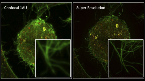
In this webinar, Stefan, Lauren, and Chunsong will discuss how to more easily create super resolution images. Olympus super resolution uses the IXplore SpinSR spinning disc confocal system to combine speed, sensitivity, and resolution for live-cell-compatible super resolution imaging. They will explain the steps involved to create super resolution images, including optical reassignment, Fourier analysis, and 3D deconvolution.

Presenters:

Stefan Marawske, Senior Sales Specialist, High-End Life Science Systems
Lauren Alvarenga, Product Manager, Life Science Microscopy
Chunsong Yan, Business Development Manager, Life Science

FAQ

Webinar FAQs | Live Cell Super-Resolution Imaging

What are the advantages of the IXplore SpinSR system over widefield-based structured illumination microscopes?

Speed is one of the major advantages of working with the IXplore SpinSR microscope. On widefield-based microscopes, the grid has to rotate many times, and the camera’s exposure time limits speed on the multiple exposures. On the IXplore SpinSR system, the spinning-disk-based optics eliminate the need for a structured illumination grid so that you can collect images faster and with fewer frames. Another advantage is that the IXplore SpinSR microscope has better depth capability—you can use a thicker specimen and still get a better image because of its confocal capabilities.

Is 3D deconvolution necessary to achieve 120 nm resolution?

The dedicated hardware optics and the Olympus Super Resolution (OSR) algorithm are the system’s key components that achieve a 120 nm resolution. However, deconvolution will further enhance the image quality and contrast.

What software runs the Olympus IXplore SpinSR microscope?

The system is run by Olympus cellSens software, which is widely used for Olympus microscopes and can instantly recognize the correct hardware. Moreover, the user interface is straightforward and intuitive. 

What kinds of detection systems are available with the Olympus IXplore SpinR microscope, and what’s the frame rate?

When imaging, the smaller the pixel, the better the resolution. With smaller pixels, you need to have better sensitivity and you also need to be careful of noise, so a Peltier-cooled scientific CMOS camera is the preferred option. We use a 2k × 2k scientific camera with a quantum efficiency of more than 80%. However, the main limitation is the sample. If you have a brighter sample, your camera’s exposure time decides the frame rate, and you won’t have to acquire multiple images to construct one super resolution image. So, if for every exposure you get one super-resolved image, you reach up to 100 frames per second. In addition, the system gives you the option to have two cameras, enabling you to image two colors simultaneously. However, the system also comes with a built-in fast emission filter that enables you to do fast sequential multicolor imaging, depending on the number of lasers and emission filters.

What types of lasers does the IXplore SpinSR system come with?

We provide all the usual lasers that conventional confocal microscopes have. We have solid state lasers that are very robust and stable and last longer than conventional gas lasers, with 405, 488, 560, and 640 as standard.

Can you use the Olympus IXplore SpinSR system for high-content screening?

Yes, absolutely. Olympus offers the advanced scanR high-content screening software. It comes with many unique features, such as the latest self-learning AI module (deep learning). It has a powerful interactive interface, can perform multilevel scanning, and is extremely versatile. Specifically concerning high-content screening, it offers exceptional speed and accuracy due to the parallel acquisition feature built into the scanR software.

Is it possible to carry out photo stimulation experiments on the IXplore SpinSR system in combination with confocal imaging?

Yes. Also, the system’s real-time controller can control older hardware through the TTL, which is much more accurate for time-dependent experiments.

Do your objective lenses have correction collars for spherical aberrations? Are they easy to adjust?

We offer A Line application-driven objectives with correction collars and a silicone immersion objective that has a long working distance. It has a correction collar, and the silicone oil more closely matches the refractive index of the sample, so you can image deeper with less aberrations. We also have some special high numerical aperture objectives, plan apochromat lenses that have an NA of 1.5 and come with a correction collar that you can set at different temperatures. So, ideally you can go up to 200 microns if you’re using a 30x silicone immersion objective.


Related Products

Super Resolution Microscope System

IXplore SpinSR

The IXplore SpinSR system is our confocal super resolution microscope optimized for 3D imaging of live cell specimens. Like the IXplore Spin system, it has a spinning disk system for fast 3D imaging while limiting phototoxicity and bleaching. However, it achieves super resolution images down to 120 nm XY and enables you to switch between widefield, confocal, and super resolution with the click of a button.

  • Sharp, clear super resolution imaging down to 120 nm XY, owing to Olympus Super Resolution (OSR)
  • Prolonged cell viability in confocal time-lapse imaging due to less phototoxicity and bleaching
  • Use two cameras simultaneously to achieve fast, two-color super-resolution imaging
  • Super resolution imaging with the world’s first plan apochromat objectives with a numerical aperture (NA) of 1.5*
* As of November 2018. According to Olympus research.

Live Cell Super Resolution Imaging: The Big Picture of Small Things

In this webinar, Stefan, Lauren, and Chunsong will discuss how to more easily create super resolution images. Olympus super resolution uses the IXplore SpinSR spinning disc confocal system to combine speed, sensitivity, and resolution for live-cell-compatible super resolution imaging. They will explain the steps involved to create super resolution images, including optical reassignment, Fourier analysis, and 3D deconvolution.

FAQ

Webinar FAQs | Live Cell Super-Resolution Imaging

What are the advantages of the IXplore SpinSR system over widefield-based structured illumination microscopes?

Speed is one of the major advantages of working with the IXplore SpinSR microscope. On widefield-based microscopes, the grid has to rotate many times, and the camera’s exposure time limits speed on the multiple exposures. On the IXplore SpinSR system, the spinning-disk-based optics eliminate the need for a structured illumination grid so that you can collect images faster and with fewer frames. Another advantage is that the IXplore SpinSR microscope has better depth capability—you can use a thicker specimen and still get a better image because of its confocal capabilities.

Is 3D deconvolution necessary to achieve 120 nm resolution?

The dedicated hardware optics and the Olympus Super Resolution (OSR) algorithm are the system’s key components that achieve a 120 nm resolution. However, deconvolution will further enhance the image quality and contrast.

What software runs the Olympus IXplore SpinSR microscope?

The system is run by Olympus cellSens software, which is widely used for Olympus microscopes and can instantly recognize the correct hardware. Moreover, the user interface is straightforward and intuitive. 

What kinds of detection systems are available with the Olympus IXplore SpinR microscope, and what’s the frame rate?

When imaging, the smaller the pixel, the better the resolution. With smaller pixels, you need to have better sensitivity and you also need to be careful of noise, so a Peltier-cooled scientific CMOS camera is the preferred option. We use a 2k × 2k scientific camera with a quantum efficiency of more than 80%. However, the main limitation is the sample. If you have a brighter sample, your camera’s exposure time decides the frame rate, and you won’t have to acquire multiple images to construct one super resolution image. So, if for every exposure you get one super-resolved image, you reach up to 100 frames per second. In addition, the system gives you the option to have two cameras, enabling you to image two colors simultaneously. However, the system also comes with a built-in fast emission filter that enables you to do fast sequential multicolor imaging, depending on the number of lasers and emission filters.

What types of lasers does the IXplore SpinSR system come with?

We provide all the usual lasers that conventional confocal microscopes have. We have solid state lasers that are very robust and stable and last longer than conventional gas lasers, with 405, 488, 560, and 640 as standard.

Can you use the Olympus IXplore SpinSR system for high-content screening?

Yes, absolutely. Olympus offers the advanced scanR high-content screening software. It comes with many unique features, such as the latest self-learning AI module (deep learning). It has a powerful interactive interface, can perform multilevel scanning, and is extremely versatile. Specifically concerning high-content screening, it offers exceptional speed and accuracy due to the parallel acquisition feature built into the scanR software.

Is it possible to carry out photo stimulation experiments on the IXplore SpinSR system in combination with confocal imaging?

Yes. Also, the system’s real-time controller can control older hardware through the TTL, which is much more accurate for time-dependent experiments.

Do your objective lenses have correction collars for spherical aberrations? Are they easy to adjust?

We offer A Line application-driven objectives with correction collars and a silicone immersion objective that has a long working distance. It has a correction collar, and the silicone oil more closely matches the refractive index of the sample, so you can image deeper with less aberrations. We also have some special high numerical aperture objectives, plan apochromat lenses that have an NA of 1.5 and come with a correction collar that you can set at different temperatures. So, ideally you can go up to 200 microns if you’re using a 30x silicone immersion objective.


Related Products

Super Resolution Microscope System

IXplore SpinSR

The IXplore SpinSR system is our confocal super resolution microscope optimized for 3D imaging of live cell specimens. Like the IXplore Spin system, it has a spinning disk system for fast 3D imaging while limiting phototoxicity and bleaching. However, it achieves super resolution images down to 120 nm XY and enables you to switch between widefield, confocal, and super resolution with the click of a button.

  • Sharp, clear super resolution imaging down to 120 nm XY, owing to Olympus Super Resolution (OSR)
  • Prolonged cell viability in confocal time-lapse imaging due to less phototoxicity and bleaching
  • Use two cameras simultaneously to achieve fast, two-color super-resolution imaging
  • Super resolution imaging with the world’s first plan apochromat objectives with a numerical aperture (NA) of 1.5*
* As of November 2018. According to Olympus research.
Experts
Chunsong Yan
Business Development Manager, Life Science
Olympus Australia & New Zealand

Chunsong is the business development manager for life science at Olympus Australia and New Zealand. He is currently responsible for confocal, multiphoton, light sheet, and slide scanning systems. He joined Olympus in 2003, working in various roles, always trying to offer the best Olympus solution to our customers.

Lauren Alvarenga
Product Manager, Life Science Microscopy

Hello. I’m Lauren Alvarenga and I work as a product manager for research imaging at Olympus where I’m currently responsible for imaging software and inverted and super-resolution microscopes. I have a B.Sc. in biomedical photographic communications from the Rochester Institute of Technology. I’ve been with Olympus since 2015, supporting the FLUOVIEW product lines in the US, Canada and Latin America.

Stefan Marawske
Senior Sales Specialist, High-End Life Science Systems
Olympus Europa

Stefan Marawske is an expert on super resolution microscopy. During his Ph.D. in the field of physical chemistry he created a home-built microscope for localization-based super resolution and particle tracking. He was fascinated by the fact that these methods are able to break the famous Abbe limit and can resolve structures that could not be identified before. He has been working with Olympus for more than 7 years and is responsible for high-end imaging systems, such as TIRF and spinning disc.

Live Cell Super Resolution Imaging: The Big Picture of Small ThingsOct 31 2024
Sorry, this page is not available in your country
Ask the Expert Sign-up

By clicking subscribe you are agreeing to our privacy policy which can be found here.

Sorry, this page is not
available in your country.

Sorry, this page is not available in your country