Evident LogoOlympus Logo

Ask the Experts

3D Assays: Intelligent Software, Insightful Analyses

3D Assays: Intelligent Software, Insightful Analyses

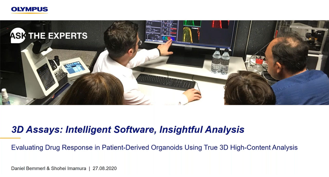
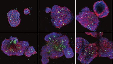
In this webinar, we discuss how to obtain the best statistical data for spheroids and microplate-based assays. These data can help you quantify the response of a 3D model to a compound and makes it easier to compare the effect of different treatments, such as varying concentrations.

Presenters:

Daniel Bemmerl, Area Manager, High-End Microscopy Systems
Shohei Imamura, Strategic Project Manager, Regenerative Medicine and Drug Discovery Solutions

FAQ

Webinar FAQs | 3D Analysis

Can NoviSight™ software be used on images obtained with other microscope brands?

Yes, it can. While NoviSight 3D cell analysis software is tailored to Olympus image files, we provide a TIFF image converter for flexibility. As long as your images are in an open format, then you can import them into the software and run the analysis.

Can NovSight software analyze 2D images?

Yes, it can. This capability is useful for broader and more standard applications.

Can widefield microscopes do 3D imaging?

It depends on the sample thickness and required outcome of your experiment. If your sample is thicker than 2–30 μm (i.e., more than 2–3 cell layers) and you want to measure single-cell level information, 3D imaging with widefield microscopy is not recommended as it is hard to visualize the depth due to light scattering.

In contrast, if your sample is less than 2-30 μm or you just want a rough estimation of the size of tissue fragments or clustered cells, such as the volume of whole organoids, it might work depending on your sample specification. Still, we would recommend applying image processing like 3D deconvolution to enhance the contrast. Please keep in mind this is still less accurate than measurement on confocal microscopes, so it is only an intermediate solution.

Can 3D applications be used in fields other than drug discovery?

Yes, definitely. We have seen a strong increase in academic research moving toward 3D data applications. For example, organoids have been studied and used in the regenerative medicine and bioengineering fields.

What microscope is best for 3D live imaging?

A two-photon microscope (also called a multiphoton microscope) is the best fit to observe a 3D live-cell sample such as a spheroid or organoid. Generally, a two-photon microscope is gentler on cells and can observe at depth within a 3D spheroid.

Does NoviSight analysis software incorporate machine learning?

The current algorithms are tailor-made for organoids and spheroids but are also flexible enough to extend to tissues. Machine learning and deep learning might make sense in the future, but for now, voxel analysis works extremely well with the traditional algorithm approach.


Related Products

3D Cell Analysis Software

NoviSight

NoviSight 3D cell analysis software provides statistical data for spheroids and 3D objects in microplate-based experiments. Use it to quantify cell activity in 3D, easily capture rare cell events, obtain accurate cell counts, and improve detection sensitivity. NoviSight software works with a range of imaging techniques, including point-scan confocal imaging, two-photon imaging, spinning disk confocal imaging, and super resolution live cell imaging.

  • Fast 3D image recognition from whole structures to subcellular features
  • Accurate statistical analysis
  • Equipped with a variety of ready-to-use default assays or easily design your own

3D Assays: Intelligent Software, Insightful Analyses

In this webinar, our experts discuss how to obtain the best statistical data for spheroids and microplate-based assays. These data can help you quantify the response of a 3D model to a compound and makes it easier to compare the effect of different treatments, such as varying concentrations.

FAQ

Webinar FAQs | 3D Analysis

Can NoviSight™ software be used on images obtained with other microscope brands?

Yes, it can. While NoviSight 3D cell analysis software is tailored to Olympus image files, we provide a TIFF image converter for flexibility. As long as your images are in an open format, then you can import them into the software and run the analysis.

Can NovSight software analyze 2D images?

Yes, it can. This capability is useful for broader and more standard applications.

Can widefield microscopes do 3D imaging?

It depends on the sample thickness and required outcome of your experiment. If your sample is thicker than 2–30 μm (i.e., more than 2–3 cell layers) and you want to measure single-cell level information, 3D imaging with widefield microscopy is not recommended as it is hard to visualize the depth due to light scattering.

In contrast, if your sample is less than 2-30 μm or you just want a rough estimation of the size of tissue fragments or clustered cells, such as the volume of whole organoids, it might work depending on your sample specification. Still, we would recommend applying image processing like 3D deconvolution to enhance the contrast. Please keep in mind this is still less accurate than measurement on confocal microscopes, so it is only an intermediate solution.

Can 3D applications be used in fields other than drug discovery?

Yes, definitely. We have seen a strong increase in academic research moving toward 3D data applications. For example, organoids have been studied and used in the regenerative medicine and bioengineering fields.

What microscope is best for 3D live imaging?

A two-photon microscope (also called a multiphoton microscope) is the best fit to observe a 3D live-cell sample such as a spheroid or organoid. Generally, a two-photon microscope is gentler on cells and can observe at depth within a 3D spheroid.

Does NoviSight analysis software incorporate machine learning?

The current algorithms are tailor-made for organoids and spheroids but are also flexible enough to extend to tissues. Machine learning and deep learning might make sense in the future, but for now, voxel analysis works extremely well with the traditional algorithm approach.


Related Products

3D Cell Analysis Software

NoviSight

NoviSight 3D cell analysis software provides statistical data for spheroids and 3D objects in microplate-based experiments. Use it to quantify cell activity in 3D, easily capture rare cell events, obtain accurate cell counts, and improve detection sensitivity. NoviSight software works with a range of imaging techniques, including point-scan confocal imaging, two-photon imaging, spinning disk confocal imaging, and super resolution live cell imaging.

  • Fast 3D image recognition from whole structures to subcellular features
  • Accurate statistical analysis
  • Equipped with a variety of ready-to-use default assays or easily design your own
Experts
Daniel Bemmerl
Area Manager, High-End Microscopy Systems

My name is Daniel Bemmerl and I’m here to help you with anything related to 3D high-content analysis and organoid imaging. I joined Olympus as an application specialist for TIRF microscopy and high-content screening and now specialize in 3D analysis software.

I studied molecular and developmental stem cell biology and quickly realized that I was more interested in the more technical aspects of research and especially in imaging itself than in the specimens I was imaging. I came to Olympus Life Science because I liked the idea of working outside of the lab and as an interface between customer and development. I'd be happy to answer any of your questions on 3D imaging and help you improve your imaging workflow to focus on your research.

Shohei Imamura
Strategic Project Manager, Regenerative Medicine and Drug Discovery Solutions

My name is Shohei Imamura. I’m a strategic project manager at Olympus. I have four years’ experience in scientific microscopy sales and seven years’ experience in product planning –especially software – and strategic project management and execution. I have a bachelor of commerce from Meiji University in Japan.

3D Assays: Intelligent Software, Insightful AnalysesNov 05 2024
Sorry, this page is not available in your country
Ask the Expert Sign-up

By clicking subscribe you are agreeing to our privacy policy which can be found here.

Sorry, this page is not
available in your country.

Sorry, this page is not available in your country